
Page 36 - 2018_12-Haematologica-web
P. 36
E. Campo et al.
20%.27,29,35,76-78 As stated earlier, minor TP53-mutant sub- clones that may be missed by Sanger sequencing also appear to carry the same unfavorable prognostic impact as clonal TP53 mutations.7,12,31,51,69
Next-generation sequencing technologies include target- ed next-generation sequencing, which has good correla- tion with Sanger sequencing in comparison stud- ies12,28,31,35,75,78 and detects low-frequency mutations below the threshold for Sanger sequencing.38,79-81 The sensitivity threshold varies depending on a number of variables, including the hardware, methods used for testing and the analytical pipeline, and should be defined by each labora-
tory using standardized criteria or equivalent medical lab- oratory standards.35,75
Reports of TP53 mutational analysis should always include the type of analysis and methodology used, the exons analyzed, the limit of detection, and coverage for next-generation sequencing (median and ≥99% mini- mum).35 Low-level TP53 mutations occurring in <10% of DNA that may be subject to further clonal selection are also identified by next-generation sequencing. Recent rec- ommendations on the methodological approaches for TP53 mutation analysis from The TP53 Network of ERIC35 concluded that the clinical importance of mutations in
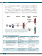
Figure 3. An example of possible clonal evolution scenarios across the course of disease in chronic lymphocytic leukemia.28,50 Genomic diversification of CLL occurs through sequential acquisition of gene mutations, represented by clones of different colors. Treatment may reduce or eliminate the incumbent clone, shifting the clonal architec- ture in favor of one or more aggressive subclones. Different therapies may preferentially provide selective advan- tages for different mutations. For example, the red circles are TP53-mutated clones, which have been selected for by chemotherapy, whereas the turquoise clones would have acquired resistance to the targeted therapy.
Table 2. Comparison of methods for the detection of TP53 aberrations.
Method Description
FISH FISH uses fluorescent DNA probes to target specific chromosomal locations within the nucleus
that can be detected by fluorescence microscopy
Sanger Sanger sequencing uses selective incorporation of sequencing chain-terminating dideoxynucleotides by DNA
polymerase during DNA replication, thereby creating sequences of various lengths, which are then separated by size to derive the DNA sequence
NGS NGS covers a range of technologies that allow high-throughput sequencing of millions
or billions of DNA strands in parallel
Genomic A technique that allows high-resolution,
arrays genome-wide screening of segmental copy number
aberrations
Advantages
• Rapid evaluation of fresh cells or paraffin-embedded interphase nuclei
• Widely used in routine clinical practice • High specificity
• Simple and widely available
• Provides direct information on mutation type • Can produce relatively long read lengths
• High specificity (~93%)
• High and customizable sensitivity
• Simultaneous analysis of large numbers
of genes
• No PCR with some platforms • Very high specificity (100%)
• Provides high resolution, genome-wide information
• Can detect genomic imbalances (deletions/amplifications) and copy-neutral loss of heterozygosity
Disadvantages References
• Can only detect genetic defects (111-114) recognized by a specific probe
• Cannot detect copy-neutral loss of heterozygosity
• Relatively time-consuming (27, 29, 35, 76-78) • Limited sensitivity (usually
approximately 10–20% of mutant
alleles)
• Limited throughput
• Upfront cost of instrumentation, although some NGS sequencers are now cheaper than capillary sequencers (for Sanger)
• High throughput needed for cost-effectiveness
• High cost
• Cannot detect balanced
rearrangements i.e. translocations, balanced insertions, inversions
(6, 27, 29, 31, 35, 76-78)
(43, 44, 48, 115-117)
FISH: fluorescence in situ hybridization; NGS: next-generation sequencing; PCR: polymerase chain reaction.
1960
haematologica | 2018; 103(12)
