
Page 35 - 2018_12-Haematologica-web
P. 35
TP53 aberrations in CLL
remaining cases with TP53 aberration carry either gene mutation(s) or sole del[17p].28,29,31,33 A TP53 mutation can be accompanied by a copy-number neutral loss of heterozy- gosity of the second TP53 allele.5,6,30,31
Clonality and clonal evolution
Individual cancer samples are genetically heterogeneous and contain clonal and subclonal populations.68,69 These populations may be in equilibrium, with the relative pro- portions of each subclone remaining stable, or may under- go evolution, with some subclones emerging as dominant.50 While most untreated CLL, and a minority of treated CLL, maintain stable clonal equilibrium, treatment may shift the architecture in favor of one or more aggres- sive subclones.50 This clonal evolution is a key feature of cancer progression and relapse, with tumors likely evolv- ing through competition and interactions between geneti- cally diverse clones (Figure 3).5 In CLL, clonal evolution after treatment or at the time of relapse has been identi- fied as ‘the rule, not the exception’.5,70 In a study by Landau et al.,5 47 out of 49 patients with CLL had clonal evolution at the time of relapse. Importantly, chemoimmunotherapy pressure is thought to lead to clonal evolution, most prominently for TP53 aberrant subclones.71
TP53 aberrations are indeed strongly associated with clonal evolution in CLL.44,72,73 TP53 aberrations are less fre- quent at diagnosis (Table 1), while 40–50% of cases with advanced or therapy-refractory CLL harbor aberrations, highlighting the need to re-assess TP53 status before each line of treatment because the clones could expand at relapse and/or during disease progression.8,10,56,74 Single or multiple minor subclones harboring TP53 mutations may
be present before therapy or may develop during relapse at any stage. These TP53-mutant minor subclones are often present at very low frequencies that may be unde- tectable by Sanger sequencing and are highly likely to expand to dominant clones under the selective pressure of chemoimmunotherapy.12,31,51
How do we test for and report TP53 aberrations? Techniques frequently used for assessing TP53 status in CLL include FISH for del(17p), Sanger sequencing, and next-generation sequencing for TP53 mutations (Table 2).27,35,74,75 As TP53 mutations are associated with a poor prognosis independently of the presence of del(17p),7 it is important to assess for TP53 mutation status using a
sequencing technique.27,35
Sequencing of the TP53 gene
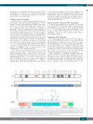
TP53 sequencing should cover exons 4–10 (correspond-
ing to the DNA binding domain at codons 100–300 and the oligomerization domain at codons 323–365) at a min- imum. Sequencing of the whole coding region (exons 2– 11) and adjacent splice sites is highly recommended using either bidirectional Sanger sequencing or next-generation sequencing, as studies of the latter have shown that vari- ants can also occur in exons outside the DNA binding domain although their frequency is low (Figure 2).35
Sanger sequencing is a widely and routinely used tech- nique to assess TP53 status in CLL in clinical practice. The technique provides a relatively simple, accessible sequenc- ing approach, but is time-consuming and lacks sensitivity for detecting minor subclones harboring TP53 mutations, with a detection limit for mutated alleles of 10–
Figure 2. TP53 gene organization and distribution of mutations by codon.63,121,122 The TP53 gene is located at the p13.1 locus on the short arm of chromosome 17 and comprises 11 exon sequences that encode for the p53 protein. While the majority of gene mutations cluster within the DNA-binding domain (codons 100–300, exons 4–8), gene mutations have been detected in almost every codon. Sequencing should, therefore, cover the DNA-binding domain and oligomerization domain as a minimum (exons 4–10), but sequencing of the whole coding region (exons 2–11) is highly recommended.
haematologica | 2018; 103(12)
1959
