
Page 23 - 2018_12-Haematologica-web
P. 23
Role of osteogenic niche in AML progression
scores cell-type-specific leukemogenic effects of various niche components. While these findings in mice offer direct evidence for osteoblast-induced leukemogenesis, emerging reports of donor cell leukemia in humans (1-5% of all post-transplant leukemia relapses), also suggest the role of an oncogenic microenvironment driving second- ary malignancy.40 Collectively, it has been increasingly recognized that genetic aberrations in the endosteal com- partment could be a key event in AML initiation and pro- gression (Figure 1).
AML induces osteogenic and osteolytic activity
Numerous AML studies have emphasized the toxicity of leukemic expansion to BM niches. AML cells have been shown to alter BM niches by competing with HSCs
for niche support, thereby affecting normal hematopoiesis.7,41,42 Whether the genomic landscape of non-hematopoietic components of BM niches changes has remained largely unexplored, and whether these alterations may drive AML initiation, progression, and resistance to chemotherapy is questionable.
Due to inconsistencies in methodology, cytogenetic analyses from different labs have led to a debate about the existence of chromosomal aberrations in leukemia patient BM-derived MSCs.10,43-45 To explore global changes induced by AML in stromal cells, our group performed a large-scale comparison of proteomic, microRNA, and gene expression profiles between AML patient-derived (AML-MSCs) and healthy donor-derived BM MSCs. We found upregulation of multiple pro-proliferative and anti-
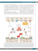
Figure 1. Osteogenic niche in hematopoietic stem cell (HSC) maintenance and leukemogenesis. Interactions between HSCs and osteogenic niche cells could hap- pen in two ways. First, HSCs stay quiescent and self-renew when they are in osteogenic niche. When they acquire mutations under physiological stress, HSCs become pre-leukemic and eventually transform into leukemia blast cells. Alternatively, genetic abnormalities in osteogenic cells in the bone marrow could induce myeloid leukemia in non-mutated or in pre-leukemic HSCs. Second, leukemic cells could induce osteogenic differentiation in mesenchymal stromal cells (MSCs), which nor- mally go through a series of differentiation steps to become fully mature osteoblasts or osteocytes. This feedback loop, involving bone remodeling, probably fuels leukemia progression. However, the extent to which acute myeloid leukemia (AML) cells induce osteogenic differentiation is not clear. BMP: bone morphogenetic pro- tein; CHIP: clonal hematopoiesis of indeterminate potential; CTGF: connective tissue growth factor; HSC: hematopoietic stem cell; LC: AML cell; OPN: osteopontin.
haematologica | 2018; 103(12)
1947
