
Page 179 - 2018_12-Haematologica-web
P. 179
Effects of Btk inhibitors on platelet activation
namely GPIb and integrin αIIbβ3. To ensure that a known degree of Btk blockade was achieved, washed platelets were incubated with ibrutinib at a concentration sufficient to fully and irreversibly inhibit Btk kinase activity (70 nM). Inhibition of Btk autophosphorylation was confirmed by
Ai
Aii
Aiii
a delay in aggregation in response to CRP (Figure 4A) and by measurement of phosphorylation (data not shown). Following incubation, platelets were reconstituted with autologous red blood cells and platelet-poor plasma and flowed over collagen at arterial shear rates. Adhesion of
Bi
Bii
Aiv
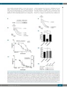
Figure 5. Patients with X-linked agammaglobulinemia, who lack Btk expression are more sensitive than healthy donors to ibrutinib inhibition of glycoprotein VI-mediated platelet aggregation. Ibrutinib, but not acalabrutinib, blocks glycoprotein VI-mediated platelet aggregation ex vivo. (A) Citrated blood was taken from XLA patients. (i) Whole cell lysates were then separated by SDS-PAGE and western blot with the polyclonal N-terminal Btk antibody. Platelet-rich plasma (PRP) from XLA patients was stim- ulated with CRP (10 mg/mL) for 180 s. (ii) A representative aggregation trace of XLA patients or healthy donor (HD). (iii) Ibrutinib dose-response curves in washed platelets of XLA patients (n=4). Healthy donor responses from Figure 2Aiii are shown as a dotted line for comparison. (iv) Whole cell lysates were then separated by SDS-PAGE and western blotted with the phosphospecific antibody to PLCg2 pY1217 (n=3). The aggregation curve for XLA patients is shown as a dotted line for comparison. (B) Patients taking ibrutinib 420 mg once daily, acalabrutinib 100 mg twice daily or a control chemotherapy regime of fludarabine (25 mg/m2 IV days 1-3), cyclophosphamide (250 mg/m2 IV days 1-3) and rituximab (375 mg/m2 IV day 1) (FCR) had citrated blood taken on day 28 of the treatment cycle (2-3 h after the dose of Btk inhibitor). PRP from this blood was then stimulated with CRP (10 mg/mL) for 180 s. (i) A representative trace. (ii) Mean and SEM from five, nine and three patients for FCR, ibrutinib and acal- abrutinib respectively. (iii) Comparison of platelet counts in PRP from all groups of patients. Statistical analysis was performed using a one-way ANOVA with the Tukey mul- tiple comparisons test, *P<0.05, ns=not significant. (iv) A representative western blot from eptifibatide (9 mM)-treated washed platelets (4×10⁸/mL) from the same patients stimulated with CRP (10 mg/mL for 180 s) followed by lysis with 5X SDS sample buffer and probed with Btk pY223 and PLCg2 pY1217 phosphospecific antibody.
Biii
Biv
haematologica | 2018; 103(12)
2103
