
Page 16 - 2018_12-Haematologica-web
P. 16
Editorials
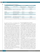
Table 1. Diagnostic biomarkers of iron-restricted erythropoiesis.*
Test
Hemoglobin g/dL
MCV MCH MCHC RDW
CHr: reticulocyte Hb content
HYPOr: % retics with Hb< 280 g/L ZPP/H: Zinc-protoporphyrin/heme ratio
Serum iron
Transferrin Transferrin saturation Ferritin
sTfR: Soluble transferrin receptor Transferrin receptor/log ferritin Hepcidin
Significance
Red blood cell indices
Iron deficiency anemia
Hb males <13 females <12
Compartment of iron in transit
Protein carrier of iron in transit
Ratio of iron to transferrin
Iron storage protein released
from storage sites
Protein derived from red cell precursors Ratio of sTfR to log ferritin
Liver derived peptide hormone
Anemia of chronic disease
Hb males <13 females <12
Hypo- micro- to normo Decreased Increased Increased
Decreased
Normal or decreased Normal or decreased Increased
Unchanged Unchanged Increased
" " " " " "
Hypochromic microcytic Decreased increased Increased
"
"
"
RBC fluorescence with Zn replacing Fe
Decreased
Increased Decreased Decreased
Increased Increased Decreased
1940
*Based on reviews by Weiss and Goodnough 2005,19 Thomas et al. 2013,20 Kiss 2015,21 Camaschella 201522 and Hempel and Bollard 2016.23 MCV: mean corpuscular volume; MCH: mean corpuscular hemoglobin; MCHC: mean corpuscular hemoglobin concentration; RDW: red cell distribution width; Hb: hemoglobin; RBC: red blood cell; Zn: Zinc; Fe: iron.
mize diagnostic biomarkers of iron deficiency anemia employing erythrocyte zinc-protoporphyrin/heme ratio (ZPP/H) and serum hepcidin measurements.
The zinc-protoporphyrin/heme ratio (ZPP/H)
The use of ZPP/H in the assessment of body iron status was reviewed in a remarkable paper by Labbe and Dewangi in 2004.3 Heme biosynthesis takes place mainly in erythroid precursor cells in the bone marrow. Iron is chelat- ed by protoporphyrin as the final reaction in the heme path- way. This reaction is catalyzed by ferrochelatase on the mitochondrial inner membrane. Iron and zinc compete for the metal binding site of ferrochelatase and when the Fe2+ substrate is insufficient, it is substituted by Zn2+, resulting in increased ZPP/H formation. Excess ZPP/H formation is a reflection of iron – zinc substrate competition for fer- rochelatase in iron-deficient erythropoiesis. ZPP/H is highly responsive to iron status even in borderline deficiency. Conversely, the decrease in ZPP/H following iron supple- mentation in preanemic states illustrates the ability of ZPP/H to respond to marginal changes in iron status. A major advantage of ZPP/H measurement is the simplicity with which it can be performed, as it requires only a portable instrument , the direct reading of fluorescence without need for any reagents, and requires minimal pro- fessional training.
The ZPP/H ratio is highly specific for iron-deficient ery- thropoiesis. However, it does not distinguish between absolute iron deficiency and iron-deficient erythropoiesis caused by anemia of chronic disease (ACD). Thus, a posi- tive test result of ZPP/H should be followed by a serum fer- ritin determination to distinguish iron deficiency from iron- deficient erythropoiesis associated with inflammation , or the toxic effect of lead exposure. Nevertheless, a ZPP/H reading within the reference range is strong evidence of adequate systemic iron supply. Indeed, as shown in a study conducted among Kenyan preschool children, when used in a screen-and-treat approach, the combination of hemo- globin concentration and whole blood ZPP/H in a single diagnostic score can be used as a rapid and convenient test- ing method to rule out iron deficiency in a substantial pro-
The study by Kanuri et al. was conducted among 4 groups (90 to 100 subjects each) of subjects selected from 2227 rural community-dwelling Indian women and preschool children. It included 90 non-anemic women, 100 non-ane- mic children, and 100 women and 100 children with IDA. Anemia was defined as hemoglobin less than 12 g/dL in women and less than 11 g/dL in children. All subjects in the normal groups had serum ferritin over 30 ng/mL and all subjects in the IDA group had serum ferritin less than 12 ng/mL and a soluble transferrin receptor (sTfR)/log ferritin index of >2. Subjects with low iron stores but normal hemoglobin were excluded from the study. The diagnostic performance of the biomarkers was estimated by analyzing receiver operating characteristic (ROC) curves to determine cut-off values with an optimal likelihood ratio>10 for IDA.
A ZPP/H ratio cut-off >90mmol/mol heme in children and >107mmol/mol heme in women was associated with a high likelihood of IDA at diagnosis (children: likelihood ratio=20.3, sensitivity 81% specificity 96%; women: likeli- hood ratio=10.8 73% specificity 93% sensitivity 73%). Hepcidin cut-off values of ≤6.8ng/mL in children and ≤4.5ng/mL in women were associated with a high likeli- hood of IDA at diagnosis (children: likelihood ratio=14.3, sensitivity 86% specificity 94%; women: likelihood ratio=16.2, sensitivity 90%, specificity 94%). The authors conclude that erythrocyte ZPP/H ratio is a valid point-of- care (POC) biomarker to diagnose IDA, and that the ZPP and hepcidin reference ranges and cut-off values identified in this study may guide clinicians to utilize these tests for the diagnosis of IDA in women and children.
In order to appreciate the significance of the data report- ed by Kanuri et al., a brief overview of our current knowl- edge on ZPP/H and hepcidin measurements for evaluating iron deficiency is presented below.
haematologica | 2018; 103(11)
