
Page 37 - 2018_11-Haematologica-web
P. 37
EMN recommendations on MM diagnosis and monitoring
The risk of pathological fractures or neurological com- plications should be assessed in patients with lytic lesions. In this regard, MRI is the preferred examination to detect spinal cord compression. If whole-body CT is not avail- able, conventional radiographs can still be used but must be interpreted with their limited sensitivity in mind. In asymptomatic patients without lytic lesions, axial MRI or whole-body MRI should be considered to assess the pres- ence of focal lesions (Grade 1B). Addition of dynamic con- trast enhancement or diffusion weighted imaging to a whole-body MRI protocol provides additional informa- tion on BM vascularization, cellularity, and composition and improves the sensitivity of MRI.50,51 Two or more focal lesions on MRI are considered as a MM-defining biomark- er.52 18F-FDG PET/CT can replace whole-body CT, if image acquisition of CT allows a detailed evaluation of the bone structures from vertex to knees, including both arms (Grade 1B).53 18F-FDG PET/CT is important to assess the presence of extramedullary disease, known to be an inde- pendent prognostic factor.54 The integration of MRI, PET- CT and whole-body CT always requires experience, inter- disciplinary consensus and reflection and needs to be cor- related with blood, urine and BM results. Finally, baseline 18F-FDG PET/CT scans enable post-treatment follow-up of hypermetabolic regions with a greater sensitivity than MRI.54,55 PET/CT is also useful in confirming MRD.56
European Myeloma Network recommendations for the diagnosis of multiple myeloma:
The initial work-up should include: complete blood count, kidney function tests, serum protein electrophoresis with immunofixation, serum albumin, β2-microglobulin, lactate dehydrogenase, C-reactive protein, calcium, serum free light chains (especially useful in the case
of light-chain multiple myeloma), 24 h protein collection with protein quantification, electrophoresis and urine immunofixation, and bone marrow (aspiration only is acceptable) studies to quantify and char- acterize abnormal plasmacytosis (Table 2). The intervals between follow-up studies depend on the response obtained and the patients’ characteristics, as proposed in Table 3 (Grade 2C). After CD138+ plasma cell sorting, fluorescence in situ hybridization analysis should include at least t(4;14) and del17p; analysis of t(14;16), 1q21 gain and del(1p32) are also recommended. In addition, bone integrity needs to be evaluated with whole-body computed tomography and/or whole-body magnetic resonance imaging (at least axial). Quantification of the level of plasma cell infiltration, serum free light chains and magnetic resonance imaging assessment are required to assess the SLIM-CRAB biomarkers that define early active multiple myeloma. At relapse, the extent of myeloma-induced bone disease should be re-evaluated, especially if the relapse occurs late after the initial diagnosis.
Staging and prognosis
Disease-specific prognostic scores
The variable outcome of MM patients depends on dif- ferences in disease biology, global disease burden and the health status of the patient. Researchers have developed clinical scoring systems in order to estimate individual prognosis. The degree of anemia, renal failure and osteol- ysis were the first disease-related prognostic biomarkers described in MM and were all included in the Salmon & Durie staging system. Subsequently, β2-microglobulin, albumin and C-reactive protein levels, and proliferative activity of MM cells were found to be additional prognos- tic factors, and albumin and β2-microglobulin levels were incorporated in the International Staging System (ISS) in
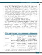
Table 3. Follow-up of multiple myeloma patients according to response and patients’ characteristics (general strength of these recommendation GRADE 2C).
Patient risk group
General myeloma population
Special patient groups
Response to prior treatment
In CR or VGPR
In PR
With biological progression
Frail patients
LC-MM and patients with renal failure
Patients with extramedullary disease
Blood and urine tests
Follow-up with blood and urine samples, initially every 1-3 months* with
gradually increasing intervals (max 6 months)
Follow-up with blood and urine samples, initially every 1-2 months* with gradually increasing intervals (max 6 months)
Regular follow-up with blood and urine samples, initially every month. Consider treatment initiation for patients with high-risk disease
Follow-up with blood and urine samples,
every 2 months; if stable increase intervals
to 3 months. Involve family doctor for follow up
Blood, SFLC and 24 h urine collection.
Follow-up with SPEP, UPEP and SFLC every month; if stable, increase intervals to 3 months
Imaging
Imaging studies should be performed when there are signs of bone disease. Consider PET/CT for high-risk patients
Imaging studies should be performed when there are signs of bone disease. Consider PET/CT for high-risk patients.
Imaging using the whole body approach is recommended.
Imaging directed to the affected region only when signs of progressive bone disease.
Imaging studies should be performed when there are signs of bone disease
PET/CT is the preferented technique for follow-up. Recommended every 6 months and obligatory in the case of progression
CR: complete response;VGPR: very good partial response; PR: partial response, PET/CT: positron emission tomography–computed tomograph; max: maximum , SPEP: serum pro- tein electrophoresis; UPEP: urine protein electrophoresis; SFLC: serum free light chain, LC-MM: light-chain multiple myeloma, 24 h: 24 hours * The monitoring intervals are gen- erally shorter for patients with high-risk disease (monthly follow-up) than for patients with standard-risk disease (every 2 to 3 months).
haematologica | 2018; 103(11)
1777
