
Page 36 - 2018_11-Haematologica-web
P. 36
J. Caers et al.
a shorter overall survival for patients with a combined del(13q14) or del(1p32). Among patients with del17p13, overall survival was shorter in those with del(1p32).33
Next-generation genome sequencing
Next-generation sequencing allows the detection of baseline clonal heterogeneity,34 clonal tiding35 and linear and branching evolution and contributes to a better under- standing of MM disease biology.36 The availability of more than 2000 sequenced MM genomes has essentially defined the genomic landscape. These data revealed a high incidence of clinically relevant genomic aberrations, including oncogenic RAS mutations, but also a number of rarer and potentially actionable lesions, such as BRAF mutations.37,38 Of note, the vast majority of available genomic data in MM is still derived from samples obtained at diagnosis and does not, therefore, necessarily reflect the situation during disease progression. In addi- tion, the clinical relevance of most mutations has not yet been determined and is undergoing investigation in large sequencing programs (CoMMpass, The Myeloma Genome Project and others).39,40 No mutation screening has yet been implemented in standard clinical workflows, but mutational analyses may help to identify potential therapeutic targets (such as BRAF mutations) and to strat- ify of patients in clinical trials.
Gene expression profiling
Based on microarrays to study mRNA expression, gene expression profiling gives a global snapshot of disease biology and may help clinicians to classify patients into separate groups with distinct outcomes. The University of Arkansas pioneered this technique to stratify MM patients
and to characterize individuals’ disease at the molecular level.41 They identified gene expression profiling patterns that allowed MM patients to be grouped in seven disease classes. Further correlation of their microarray results with survival data of individual patients identified a list of 70 genes (GEP70) that had strong prognostic information.41 Similarly, the HOVON group identified a 92-gene signa- ture (termed SKY92), based on the gene expression profil- ing results of the Hovon-65 trial.42,43 Other gene expression profiling-based risk models have been developed, such as the IFM-15 and MRC-IX-6 gene signatures44,45 Although not routinely determined in the majority of laboratories within or outside Europe, both the GEP70 and the SKY92 profiles are commercially available.
Imaging
Traditionally, osteolytic bone disease was investigated by conventional skeletal radiography. The 2014 IMWG disease criteria also considered small osteolytic lesions (≥5 mm), detected by computed tomography (CT) or com- bined 18F-fluorodeoxyglucose (18F-FDG) positron emission tomography (PET/CT) as symptoms of myeloma-induced bone disease.3 Taking into account these definitions, in 2015 the EMN proposed a relevant algorithm for guiding the choice of imaging technique.46,47 Different European centers have integrated CT into their diagnostic work-up based on its superior sensitivity and its ease of operation This choice was supported by the recent implementation as a national standard of care in the diagnostic workup of patients with suspected MM in the UK and elsewhere.48 Whole-body CT has also been integrated into the diagnos- tic work-up of the European Society of Medical Oncology49 and the upcoming IMWG guidelines.
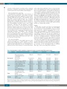
Table 2. Recommendations on further examinations at diagnosis, for response assessment, during follow-up and at relapse.
Diagnostic site
Bone marrow
Blood
Urine
Imaging
Tool
BM cytology and biopsy to confirm plasmacytosis and monoclonality
Flow cytometry
Cytogenetics Obligatory
Diagnosis
Obligatory
At response
Obligatory*
Optional
Not required
Not required Obligatory Obligatory Recommended *** Obligatory Obligatory Obligatory Obligatory Recommended
Obligatory Recommended†
Not required Optional††† Not required
At follow-up
Not required
Not required
Not required
Not required Obligatory Obligatory
At relapse
Obligatory**
Optional
Optional
Not required Obligatory Obligatory Recommended *** Obligatory Obligatory Obligatory Obligatory Obligatory
Obligatory Recommended†
Recommended Optional Optional
Recommended
Advanced techniques: GEP, NGS Blood count and blood smear Serum electrophoresis and IF Serum free light chain
Serum immunoglobulin levels Renal and liver function tests Calcium
Lactate dehydrogenase Albumin, β2-microglobulin
Urine sample to check for proteinuria and Bence-Jones proteins
24 h urine collection
Low dose whole-body CT PET/CT
Whole-body MRI
Optional Obligatory Obligatory
Recommended Obligatory Obligatory Obligatory Obligatory Obligatory
***
Recommended Obligatory Obligatory Obligatory Obligatory
***
Obligatory Recommended†
Recommended†† Optional Optional
Recommended Obligatory
Recommended†
When symptomatic When symptomatic When symptomatic
BM: bone marrow; GEP: gene expression profiling; IF: immunofixation; NGS: next generation sequencing; CT: computed tomography; PET: positron emission tomography; MRI: magnetic resonance imaging; *Obligatory for patients in complete response. **Obligatory for patients with light chain escape, oligosecretory disease, *** SFLC monitoring is obligatory for patients with light-chain disease.†Obligatory in the case of proteinuria.††Obligatory when radiographs do not show osteolytic lesions †††PET/CT is required for con- firmation of minimal residual disease negativity.
1776
haematologica | 2018; 103(11)
