
Page 127 - 2018_11-Haematologica-web
P. 127
Targeting FLT3-ITD in AML
ed as, compared to primary FLT3-ITD AML cells with 80% blast cells, FLT3-ITD AML cell lines with 100% blast cells are more susceptible to midostaurin (Figure 2A), and less susceptible to NVP-HDM201 (Figure 2B).
With respect to midostaurin, FLT3-ITD/TP53wt NK- AML cells were distinctly more susceptible than FLT3- ITD/TP53wt cells. Patient derived AML blast cells charac- terized by FLT3-ITD/TP53wt/NPM1wt were susceptible to midostaurin with a median loss of 30% viability after treatment with 500nM PKC412.
FLT3-ITD/TP53wt/NPM1wt cells with 11q23/MLL abnormalities, MOLM-13 (t(9;11) and MV4-11 (t(4;11) lost 60% cell viability within 24 hours when treated with 500nM midostaurin. All other AML cells including
FLT3-ITD/TP53wt/ NPM1mut, FLT3wt and TP53mut cells were less susceptible to midostaurin with 0-10% reduced viability at 500nM midostaurin (Figure 2A).
With respect to NVP-HDM201, we observed that the same NK-AML blast cells characterized by FLT3- ITD/TP53wt/NPM1wt and sensitive to midostaurin were most susceptible to the MDM2 inhibitor NVP-HDM201, with a median loss of 45% viability within 24 hours at 100nM NVP-HDM201. MOLM-13 and MV4-11 cells were less susceptible with a loss of 20% viability at 100nM NVP-HDM201. FLT3-ITD/TP53wt/NPM1wt cells responded with a median 20% loss of viability and FLT3- ITD/TP53wt/NPM1wt with median 10% loss of viability at 100nM NVP-HDM201. All other AML cells including
AB
CD
EF
GH
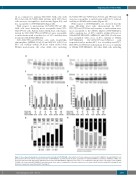
Figure 4. Dose-dependent induction of apoptosis and cell death in FLT3-ITD AML cells. Induction of tumor suppressor protein p53 in MV4-11 (A) and MOLM-13 cells (B) treated for 24 hours with the indicated amounts of NVP-HDM201 and midostaurin. Relative quantitation of CDKN1A mRNA (C) and MCL-1 mRNA (D) in AML cells treated for 24 hours with midostaurin (PKC412) (black bars) and NVP-HDM201 (light grey bars) alone or in combination (dark grey bars). Cytometric assays in MV4- 11 AML cells treated with NVP-HDM201 and midostaurin alone and in combination to measure induction of cell death (subG1 fraction) using DAPI staining (E) and induction of apoptosis using AnnexinV/PI staining (F).
haematologica | 2018; 103(11)
1867
