
Page 178 - 2018_10-Haematologica-web
P. 178
M. Rijkers et al.
ments of WIM8E5, SN607D8 and SN230G6 were generat- ed (Online Supplementary Figure S4A). The lack of an Fc-tail (Online Supplementary Figure S4B) and binding (Online Supplementary Figure S4C) of the F(ab’)2 fragments to HLA molecules on platelets was confirmed. Staining with anti- IgG directed to the Fc-tail of human IgG was negative for F(ab’)2 fragments and positive for IgG. For WIM8E5, F(ab’)2
A
binding was lower than that of the corresponding IgG, but significant binding was still observed (Online Supplementary Figure S4C). None of these F(ab’)2 fragments induced CD62P membrane exposure (Online Supplementary Figure S4D), indicating that crosslinking of an HLA molecule and FcγRIIa by an intact anti-HLA IgG is crucial to induce platelet activation.
B
C
DE
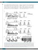
Figure 1. HLA monoclonal antibodies induce platelet α-granule release. (A) Platelets were matched for HLA type with the specificity of eight HLA monoclonal anti- bodies (mAbs) directed at different epitopes. Mean fluorescent intensity (MFI) upon staining with anti-human IgG was measured with flow cytometry for the control (buffer only, no HLA antibodies) and 2.5 mg/mL of the HLA mAbs. Right panel: representative flow cytometry plot of 10 mg/mL SN607D8 with a not matching donor and a matching donor. (B,C) CD62P surface expression of platelets incubated with 2.5 mg/mL (B) or 10 mg/mL (C) HLA mAbs compared to control (buffer only). Representative flow cytometry plots of WIM8E5, SN607D8 and SN230G6. (D) VWF release in platelet supernatant upon incubation with HLA mAbs WIM8E5, SN607D8 and SN230G6 measured by enzyme-linked immunosorbent assay. (E) Representative western blot of SPARC release in platelet supernatant upon incuba- tion with HLA mAbs WIM8E5, SN607D8 and SN230G6. Paired t-tests (A, B and C) or paired ANOVA with the Tukey multiple comparison test (D). Each line represents a separate experiment with a separate donor (A, B and C). Mean ± SD (D). *P<0.05, **P<0.01, ***P<0.005, ****P<0.001.
1744
haematologica | 2018; 103(10)
