
Page 126 - 2018_10-Haematologica-web
P. 126
A. Liu et al.
expression is expressed by more mature Mks.18 Hence, CD41a+/CD42b– represents immature Mks, and CD41a+/CD42b+ represents more mature Mks. Flow cyto- metric analyses showed that the proportion of immature CD41a+/CD42b– cells was significantly increased in the presence of IMiDs (day 7: vehicle 15.8%, LEN 23.0 %, POM 22.9%; day 10: vehicle 32.1%, LEN 50.6%, POM 51.0%; day 14: vehicle 49.7%, LEN 64.7%, POM 66.0%; P<0.05). This difference was visible as soon as day 7 of culture and was maintained throughout the entire culture period (Figure 2D). To confirm the immature and dysplas- tic morphology, we performed transmission electron microscopy (TEM) after 10 days of IMiD treatment. Mks were identified by CD61-microbead labeling. Again, com- pared with vehicle, IMiD-treated Mks exhibited immature
features, including decreased size, less cytoplasm, and a heterogeneously dilated and abnormally distributed demarcation membrane system (DMS) in the cytoplasm. (Figure 2E)
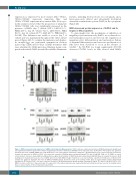
IMiD decreased protein expression of GATA1 and its targets in Mk progenitors
To gain insight into the mechanism of inhibition of megakaryocytic maturation by IMiDs, we evaluated sev- eral transcription factors involved in the regulation of megakaryocytic differentiation and maturation. Failure of terminal differentiation and excessive proliferation of Mks have been described to occur in the absence of GATA1.19 By RT-PCR we found significantly (P<0.05) decreased GATA1 levels in Mks treated with IMiDs
AB
C
D
Figure 3. IMiD decreased protein expression of GATA1 and its targets in Mk progenitors. CD34+ cells were cultured in serum-free HPGM hematopoietic growth medi- um supplemented with 10 ng/mL TPO to initiate megakaryopoiesis with or without 10 mM IMiDs for the indicated times. (A) Real-time PCR analysis was applied to determine the levels of GATA1 mRNA. The result shown here is one representative experiment of triplicates. (B) Immunofluorescence microscopy was performed to examine the expression of GATA1 on CD34+ cells cultured in serum-free HPGM medium containing 10ng/mL TPO with 10 mM LEN, POM or 0.1% DMSO as vehicle control at day 6. Cells were stained with an antibody that recognize the N terminus of GATA1 (red) and counterstained with DAPI (blue) to visualize the nucleus. Both stainings were merged and revealed a loss of GATA1 when treated with IMiDs. Images shown here is one representative experiment of triplicates. (C) Western blot analysis was applied to determine the levels of GATA1 and IKZF1 protein. β-actin was used as the loading control. Data are representative of three independent experiments. (D) Purified CD34+ cells were cultured in serum-free HPGM hematopoietic growth medium containing 10 ng/mL TPO with 10 μM LEN, POM or 0.1% DMSO as vehicle control for 6 days and 9 days. The expression of the indicated proteins was analyzed by Western blotting. b-actin was used as the loading control. Data are representative of three independent experiments.
1692
haematologica | 2018; 103(10)
