
Page 125 - 2018_10-Haematologica-web
P. 125
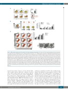
IMiDs block megakaryocytic maturation
A
B
D
C
*P<0.05 **P<0.001
Figure 2. IMiDs inhibit megakaryocytic differentiation by blocking endomitosis and maturation. (A) Upper panel: Flow cytometry analysis revealed a strong induc- tion of CD34+CD38–cells (early progenitors) after 14 days of treatment with POM +/- TPO compared to vehicle. Lower panel: Early CD34+CD38– progenitors were fur- ther analyzed for CD33+ and CD41+ expressions. Lower right panel: The mean fluorescence intensity (MFI) of CD33 in these CD34+CD38– immature hybrid cells dra- matically increased with TPO. (B) In long-term cultures, CD34+ cells were maintained for up to 4 months and multicolor flow cytometry identified 2 cell populations: CD45+34+33+11b+41+61+ hybrid cells (6.6%) and a more mature population of CD45+34-41+61+ cells (86.6%). Without POM, the control cultured cells could be main- tained only for up to 3 weeks and were therefore not available for comparison. Data are from one experiment. (C) Purified CD34+ cells were cultured in serum-free HPGM hematopoietic growth medium with DMSO or IMiDs. After culturing for 14 days, CD34+ cells from each group (vehicle, LEN and POM) were selected by immuno- magnetic beads and were plated in MethoCult for colony formation assays (with either vehicle, LEN or POM). Data are from one experiment with colonies quantified in triplicate wells. *P<0.05 compared to Vehicle; **P<0.001 compared to Vehicle. Data were compared by one-way ANOVA with Bonferroni post-test. (D) CD34+ cells were cultured in serum-free HPGM hematopoietic growth medium with TPO to induce megakaryopoiesis with or without LEN, POM or DMSO as vehicle. After 7, 10 and 14 days, flow cytometry analysis showed an increase of immature Mks (CD41a+/CD42b–), while more mature Mks (CD41a+/CD42b+) decreased with IMIDs treat- ment. The result shown here is one representative experiment of triplicates. (E) Morphology of Mks, derived from CD34+ cells grown in serum-free HPGM hematopoi- etic growth medium with TPO with or without IMiDs at day 7 and 10 of culture. Electron micrographs of three characteristic Mks from different groups (vehicle, LEN and POM) are shown. Mks were identified by CD61+ magnetic bead labeling. Mks in LEN and POM group show features of immaturity, including scant cytoplasm, large nuclei, and a minimal demarcation membrane system. Images are from one experiment representative of two independent experiments each of which included triplicates.
renewal beyond direct exposure, we first cultured CD34+ cells in liquid cultures under the conditions described above. After 14 days, CD34+ cells were selected, and sub- jected to colony formation assays without adding IMiDs. Even without the continued presence of IMiDs, total colony formation was still significantly increased [vehicle 120, LEN 256 (P<0.05), POM 270 (P<0.001)]. In accor- dance with prior data,17 we again observed a shift to myeloid lineage commitment with increased myeloid colonies (CFU-G, CFU-GM and CFU-GEMM) at the expense of erythroid colonies (P<0.05; Figure 2C). These data suggested that IMiDs persistently affect self-renewal
and lineage commitment of CD34+ cells, resulting in main- tenance and expansion of the progenitor cell pool. Our finding showed that IMiDs-induced development of megakaryocytic precursors appears to contrast with the thrombocytopenia in patients caused by IMiDs. Since our previous studies showed that IMiDs are not directly toxic to bone marrow hematopoietic cells,16 we hypothesized that these effects are induced by maturational arrest. To investigate the effects of IMiDs on the maturation of Mks, we evaluated different megakaryocytic cell populations by flow cytometry. CD41a is a lineage marker of Mks throughout all stages of differentiation, whereas CD42b
haematologica | 2018; 103(10)
1691
