
Page 69 - 2018_09-Mondo
P. 69
TGFβ1-mediated inhibition of MDS- and AML-derived MSC
ing within the bone marrow microenvironment influ- ences the stromal compartment in addition to HSPC, thereby contributing to insufficient hematopoiesis.
Ingenuity pathway analysis and GSEA predicted that the inhibitory cytokine TGFβ1 (but not TGFβ2, data not shown) is an upstream regulator of the observed functional and molecular alterations in MDS- and AML-derived MSC. Again, this confirms and expands the results from Chen et al., who recently identified signatures related to TGFβ signaling in primary MSC from patients with low- risk MDS.10 This molecular similarity between our cul- ture-expanded MSC and the non-expanded, FACS-sorted MSC reported by Chen et al. also indicates that the most relevant pathophysiological mechanisms seem to be well preserved even after culture. Apart from its pathophysio- logical relevance this also has important methodological implications. The use of non-expanded FACS-sorted MSC does not allow subsequent direct functional testing of candidate genes because of the low number of cells, but also requires culture or cell lines.10 In contrast, we were able to test the effects of TGFβ1 on stromal cell functionality in the same culture system previously used to isolate and characterize MDS- and AML-derived MSC. This methodological issue should ideally be further inves- tigated by a direct comparison of matched FACS-sorted,
A
B
C
unexpanded and culture-propagated MSC from the same individual patients.
We did not detect any relevant effects on MSC func- tions after exposure to TNFa (data not shown). Considering the limited clinical efficacy of TNFa block- ade by etanercept and infliximab in MDS patients,34-36 we believe that TNFa plays, at most, a minor role. In con- trast, exposure to TGFβ1 suppressed growth and osteogenic differentiation of healthy MSC and induced changes in the expression of candidate genes (PITX2, HOXB6, TBX15, Kit-ligand, Angiopoietin-1, and Jagged1). Together with impaired stromal hematopoietic support, the phenotype of healthy MSC therefore resembles that of primary MDS- and AML-derived MSC after exposure to TGFβ1.12,13 The inhibitory effect of TGFβ1 on normal HSPC is well documented.37 In addition, overactivation of TGFβ signaling in CD34+ HSPC has been shown to be an important mechanism mediating hematopoietic insuffi- ciency in MDS.23,38,39 Growth differentiation factor-11, another ligand of the TGFβ superfamily, inhibits matura- tion of erythroid precursors and elevated levels of this factor have been found in patients with MDS, implicating it in the development of anemia.40 In addition to suppres- sion of HSPC and erythroid progenitors, our data from RNA sequencing and functional experiments show for
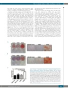
Figure 6. Blockage of transforming growth factor β1 signaling by SD-208 restores the osteogenic differentiation capacity of myelodysplastic syndrome- and acute myeloid leukemia-derived mesenchymal stromal cells. Osteogenic differentiation of MDS- and AML- derived MSC was induced and visualized by Alizarin red staining. SD-208 (0.5 μM) was added to the osteogenic medium during this procedure. (A) Overview of a representative experiment investigating MDS-derived MSC (n=5, MDS patients n. 1, 2, 7, 12, 13). Representative micro- graphs of MDS-derived MSC after exposure to SD-208 with scale bars indicating 100 μm. (B) Overview of a representative experiment investigating AML-derived MSC (n=4, AML patients n. 1, 5, 12, 13). Representative micrographs of AML-derived MSC after exposure to SD-208 with scale bars indicating 100 μm. (C) Differences in osteogenic differentiation between patient- derived MSC (black bars, MDS n=5; AML n=4) either native or treated with SD-208 (white bar) were quantified by a previously described score.12 In detail, differentiation was graded accord- ing to microscopic analysis of staining intensity as follows: 0 = absent; 1 = weak; 2 = moderate; 3 = intensive. For all experiments results are expressed as mean ± SEM. Asterisks indicate P- values **P<0.01, ***P<0.001.
haematologica | 2018; 103(9)
1469
