
Page 67 - 2018_09-Mondo
P. 67
TGFβ1-mediated inhibition of MDS- and AML-derived MSC
Transforming growth factor β1 impairs stromal hematopoietic support function
We have previously described the deregulation of Jagged1, Angiopoietin-1 and Kit-ligand, three signaling molecules physiologically involved in the regulation of HSPC, in patient-derived MSC.12,13 We now examined whether TGFβ1 can also induce alterations of these fac- tors and of three candidate cytokines (CCL26, SPP1 and LIF) that were shown to be deregulated in primary patient-derived MSC. Indeed, quantitative PCR and flow cytometry detected an expression pattern (Figure 5A,B, Online Supplementary Figures S10 and S11) that is congru- ent with the expression previously detected in primary MDS- and AML-derived MSC.12,13 Again, these effects were reversible by adding SD-208 (Figure 5A,B).
In light of the profound molecular alterations that were induced by TGFβ1 in healthy MSC, we were further interested in whether TGFβ1 also suppressed stromal hematopoietic support. We therefore used the established long-term culture-initiating cell (LTC-IC) assay and cul- tured healthy CD34+ HSPC on human MSC feeder layers which had previously been incubated with TGFβ1 or with TGFβ1 together with SD-208 for 28 days. As indi- cated in Figure 5C, TGFβ1 significantly inhibited the abil- ity of healthy MSC to support CD34+ HSPC in the LTC- IC assay, an effect that could be reversed by inhibiting TGFβ1 signaling through SD-208. In summary, TGFβ1
induces a phenotype and functional deficits recapitulating those observed in primary MSC obtained from patients with MDS or AML. Our data thus strongly support the hypothesis derived from RNA sequencing data analysis, namely that TGFβ1 contributes significantly to the func- tional inhibition of MSC in these two myeloid malignan- cies.
Inhibition of transforming growth factor β1 signaling restores osteogenic differentiation and hematopoietic support capacity of myelodysplastic syndrome-
and acute myeloid leukemia-derived mesenchymal stromal cells
In a final set of experiments we directly addressed the impact of TGFβ1 on MSC functionality in human MDS and AML. To abrogate the potential influence of TGFβ1 on MSC functions we again used SD-208. This inhibitor had already been demonstrated to abolish TGFβ signaling in the above-mentioned experiments, as well as previous- ly in MDS-derived CD34+ cells.12,13 Given the substantial inhibition of osteogenic differentiation in primary patient-derived MSC, we focused on the potential effects of TGFβ1 on osteogenesis. For this purpose we cultured MSC, freshly isolated from the bone marrow of patients with MDS and AML, in the presence of SD-208 and doc- umented that osteogenic differentiation capacity could be significantly rescued compared to that occurring in the
ABC
DEF
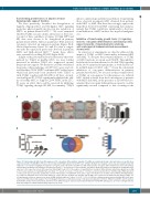
Figure 4. Transforming growth factor β1 suppresses the osteogenic differentiation capacity of healthy mesenchymal stromal cells and induces a specific gene expression profile. Healthy MSC (n=6) were pre-incubated with TGFβ1 and/or SD-208 for up to 28 days. Medium was changed every 3 days and supplemented with TGFβ1 at a concentration ranging from 5 ng/mL to 10 ng/mL and/or SD-208 (0.25 μM to 0.5 μM). Subsequently, osteogenic differentiation was induced for 14 days and visualized by Alizarin red staining as described previously.12,13 (A) Overview of a representative experiment. (B) Representative micrographs of healthy MSC after exposure to the respective factors with scale bars indicating 100 μm. (C) For the purpose of quantification, osteogenic differentiation capacity was graded according to microscopic analysis of staining intensity as follows: 0 = absent; 1 = weak; 2 = moderate; 3 = intensive as previously described.12 (D) Messenger RNA expression of osteocalcin was measured by quantitative real-time PCR analysis of healthy MSC (n=5) after 3 days of incubation. HC: healthy control; DMSO: dimethylsulfoxide; w/o TGFβ1: healthy MSC were treated with TGFβ1 for 3 days, then the medium was changed and the MSC were cultured for 3 additional days without TGFβ1. For all experiments results are expressed as mean ± SEM. Asterisks indicate P-values *P<0.05, **P<0.01. (E) After a 28-day incubation period healthy MSC (n=2) were subjected to RNA sequencing analysis. The Venn diagram illustrates a substantial overlap of 312 genes deregulated in both the TGFβ1-treated MSC and the patient- derived MSC. (F) Bar charts demonstrate that most of the differential gene expression provoked by TGFβ1 in healthy MSC (1812 genes, q<0.05) is abrogated by SD- 208 (only 58 genes remained differentially expressed, q<0.05).
haematologica | 2018; 103(9)
1467
