
Page 170 - 2018_09-Mondo
P. 170
J. Qiao et al.
AB
C
C
D(i)
D(ii)
E(i) E(ii)
E(iii)
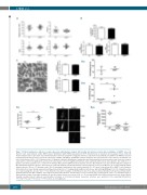
Figure 1. Platelet parameters, adhesion receptor expression, ultrastructure analysis, tail bleeding and arterial occlusion time in wild-type or NLRP3-/- mice. (A) Platelet count, mean platelet volume (MPV), platelet distribution width (PDW) and plateletcrit (PCT) determined by an automatic blood analyzer (mean ± SE). (B) Platelet aIIbβ3 surface expression was determined by flow cytometry using FITC-conjugated anti-mouse aIIβ monoclonal antibody; meanwhile total RNA was isolated from washed platelets in order to assess the expression of GPIba and GPVI by quantitative real-time PCR. Data are represented as a ratio relative to an internal con- trol (β-actin) (mean ± SE, n = 5-7) (Student t-test). (C) Analysis of platelet ultrastructure (a-granules and dense granules) by electron microscopy. Scale bar: 2 μm for upper panel (x 15,000 magnification) and 1 μm for lower panel (x 30,000 magnification). Black arrow: a-granule; white arrow: dense granule. The numbers of a- granules and dense granules were counted in 60 wild-type (WT) and 60 NLRP3 knockout platelets (mean ± SE) (Student t-test). (D)-i Tail bleeding time analysis of WT and thrombocytopenic (TP) mice after injection of anti-aIIb antibody, followed by infusion of donor platelets (1 x 10⁸) from WT mice at 9 h after antibody admin- istration (mean ± SD, n= 6-7) (Student t-test). (D)-ii To investigate hemostasis in vivo, washed platelets (1 x 10⁸) were isolated from WT or NLRP3-/- mice and then infused into mice made thrombocytopenic by intraperitoneal injection of 0.1 mg/kg (body weight) rat anti-mouse aIIb antibody (MWReg 30) for 9 h, followed by analy- sis of tail bleeding time (mean ± SD, n = 5-6) (Student t-test). (E) For analysis of thrombosis in vivo, FeCl3-induced arterial thrombus formation was initiated after washed platelets from WT or NLRP3-/- mice had been infused into WT mice and the time to vessel occlusion was recorded (mean ± SD, n = 9) (Student t-test) (i). Representative image of thrombus formation (ii) and the relative fluorescence (mean ± SD, n = 5) (one-way ANOVA) (iii) at different time points are shown. Movies showing real-time platelet adhesion and thrombus formation in FeCl3-induced injured mesenteric arterioles were provided in the supporting information. *P<0.05;**P<0.01; ***P<0.001; ****P<0.0001. ns: not significant.
1570
haematologica | 2018; 103(9)
