
Page 160 - Haematologica August 2018
P. 160
A. Roberto et al.
after the transplant (Figure 1C,D). Supervised hierarchical clustering revealed that in healthy donors uCD56dim NK cells have a gene expression similar to that of cCD56dim cells and different from that of cCD56bright NK cells. These results were somewhat expected considering that both uCD56dim and cCD56dim NK cells share a high degree of cytotoxicity in physiological conditions.2 Although show- ing distinct gene expression profiles, cCD56bright and uCD56dim NK cells from hHSCT recipients group together and are more similar to cCD56dim than cCD56bright NK cell subsets from healthy donors (Figure 4B).
We then compared the fold change (FC) of the 3072 dif-
ferentially expressed genes between uCD56dim NK cells
from hHSCT patients and cCD56bright (FCx) or cCD56dim
(FCy) NK cells from healthy donors (Figure 4C). Among
those transcripts of uCD56dim NK cells from hHSCT
patients similarly expressed in cCD56dim NK cells from
healthy donors (FCy (log2) < |1|) but differently modulated
in cCD56bright NK cells from healthy donors (FCx (log2) >
|1|), we found a downregulation of CCR7, interleukin (IL)-
from healthy donors (FCx (log2) < |1|) but differently mod- ulated in comparison with cCD56dim NK cells from healthy donors (FCy (log2) > |1|) we found a downregulation of the maturation marker KLRG1 (Figure 4C3), of CD160 and GRZ-M (Figure 4C3).
These transcriptional profiles parallel our flow cytome- try data and confirmed our working hypothesis postulat- ing that uCD56dim NK cells present at high frequencies early after hHSCT are not NK cell precursors, but rather represent lymphocytes in a later stage of differentiation whose gene expression profile is intermediate between cCD56bright and cCD56dim NK cells.
Highly proliferating NKp46neg-low/uCD56dim cells can generate NKp46pos/cCD56bright NK cells
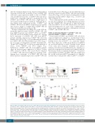
Donor-derived uCD56dim and cCD56bright NK cell subsets expressed high levels of Ki-67 (i.e., proliferating) at three and four weeks after hHSCT, while their counterparts in healthy donors were all Ki-67neg (i.e., quiescent) (Figure 5A). These high rates of NK cell proliferation in hHSCT are associated with the so-called “cytokine storm” which occurs early after allogeneic transplant and induces immune cell activation and differentiation to rapidly recu- perate the recipients from the previously induced and life- threatening condition of immunodeficiency.25,31 The prefer- ential expansion of Ki67pos/uCD56dim NK cells starting from the second week after hHSCT prompted us to hypothe- size that they could generate either cCD56bright or cCD56dim
7R and TCF7 (Figure 4C ) as well as an upmodulation of 1
CCL3, CCL4, PRDM1 and IFN-γ (Figure 4C ). 2
Furthermore, uCD56dim NK cells from hHSCT patients show an increased gene expression of NKp30, Perforin (PRF), Granzyme (GRZ-B), GRZ-A and GRZ-H compared to cCD56bright NK cells from healthy donors (Figure 4C2). Among those transcripts of uCD56dim NK cells from hHSCT patients similarly expressed in cCD56bright NK cells
AB
CD
Figure 5. FACS-sorted uCD56dim NK cells generate cCD56br NK cells under IL-15 and IL-18 stimulation. (A) Summary statistical graph showing the expression of Ki67 on cCD56bright (blue), cCD56dim (black) and uCD56dim (red) from HSC healthy donors (HDs) and their recipients after three and four weeks from hHSCT. (B) Representative example from a HD of flow cytometry dot plots showing the purity of fluorescence-activated cell sorting (FACS)-sorted cCD56bright (blue), cCD56dim (black) and uCD56dim (red) (left column) natural killer (NK) cell subsets. Highly pure and FACS-sorted NK cell subsets are overlaid with the phenotype of purified CD3neg/CD20neg NK cells expressing CD56 and CD16 (gray). (C) Summary statistical graphs showing the proliferation index of FACS-sorted cCD56bright (blue) and uCD56dim (red) NK cell subsets from six HDs at four, eight and 14 days of culture with interleukin (IL)-15+ IL-18. (D) Summary statistical graph showing the kinetic of CFSE-diluting (CFSEdil) cCD56bright (blue), cCD56dim (black) and uCD56dim (red) NK cell subsets generated from FACS-sorted cCD56bright (upper panel) and uCD56dim (lower panel) from seven HDs. No data are available for the cCD56dim NK cells, as they were not proliferating in response to IL-15 and IL-18. Data are expressed as means ± S.D. *P<0.05; **P<0.01; ***P<0.001.
1396
haematologica | 2018; 103(8)
