
Page 137 - Haematologica August 2018
P. 137
Therapeutic effects of chidamide on myeloma-associated bone disease
revealed greater myeloma-associated bone loss in the vehicle group as compared with the chidamide-treated group (Figure 5D). Moreover, based on the bone morpho- metric parameters, chidamide increased the trabecular number (**P<0.01) and bone volume density (P=0.0755) and reduced trabecular separation (**P<0.01) compared to the control group (Figure 5E).
Chidamide inhibits osteoclastogenesis and bone resorption in vitro
As the in vivo study revealed that chidamide exerts anti- myeloma effects, we investigated the effect of chidamide on the formation and function of OCs of human origin. The expression of DUSP1, c-fos, NFATc1 and HDAC10 increased during RANKL-induced OC formation (Figure 6A). We then tested several key factors and signaling path-
ways that mediate osteoclastogenesis in order to identify the mechanisms underlying the aforementioned effects. The levels of p-p38, p-ERK1/2, p-JNK and p-AKT were also reduced, indicating that chidamide suppressed the classical pathways of OC activation. Cathepsin K, c-fos, HDAC10 and NFATc1 expression levels were all down- regulated after chidamide treatment in a dose-dependent manner. In addition, acetylation of H3K9, H3K18 and H4K8 was increased after chidamide treatment (Figure 6B). PBMCs cultured in osteoclastogenic medium were incubated with different concentrations of chidamide to evaluate its effects on OC formation. At a low concentra- tion (0-1μM) of the drug, the number of TRAP+ multinu- cleated cells derived from healthy donors was reduced, but the density of cells was not significantly reduced (**P<0.01, Figure 6C,D). Additionally, F-actin ring forma-
A
B
B
C
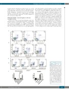
Figure 3. Chidamide overcame the resistance conferred by chi-
damide-pretreated OCs BMSCs. (A) and (C) H929 cells were co-cultured with OCs and chi- damide-pretreated OCs for 48h or with 6μM chidamide, and cell apoptosis was detected by flow cytometry after staining with Annexin V and PI. (B) and (C) H929 cells were co-cultured with BMSCs or with 6μM chidamide for 48h, and cell apoptosis was detected by flow cytometry after staining with Annexin V and PI (H929+chidamide vs. H929+OCs+chidamide, ***P<0.001; H929+chidamide vs. H929+OCs(chida)+chidamide, P>0.05, non-significant; H929+chi- damide vs. H929+BMSCs+chi- damide, P>0.05 non-significant). PI: propidium iodide; OC: osteo- clasts; BMSC: bone marrow stromal cell.
and
haematologica | 2018; 103(8)
1373
