
Page 135 - Haematologica August 2018
P. 135
Therapeutic effects of chidamide on myeloma-associated bone disease
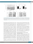
Figure 1. Chidamide exhibits HDAC inhibitory activity in MM cell lines. (A) Western blotting was used to detect the protein levels of HDAC1, HDAC2, HDAC3 and HDAC10. (B) HDAC activity assay showed that chidamide could significantly inhibit HDAC activity (Both in ARP-1 and RPMI-8226, ***P<0.001). (C) Western blot analysis of H3 acetylation at Lys18 and Lys9 and H4 acetylation at Lys8. H3 and H4 expression levels were used as loading controls. (D) Western blot analysis of HDAC1, HDAC2, HDAC3, and HDAC10 levels. Acetylation of α-tubulin at K40 was also examined, and a-tubulin expression was used as the loading control. HDAC: histone deacetylase.
A
B
CD
and propidium iodide [PI] staining) showed that chi-
damide significantly induced MM cell apoptosis at 48h
and 72h (Figure 2A and Online Supplementary Figure S1B).
donors were incubated with 4μM chidamide for 48h to examine the cytotoxicity of chidamide toward non-tumor cells. Although approximately 10% of monocytes under- went apoptosis, the numbers of apoptosis cells were only slightly higher than the vehicle group and far less than MM cells (Online Supplementary Figure 2B).
Furthermore, we investigated the cytotoxicity of chi- damide in the presence of the BM microenvironment. For this purpose, H929 cells were co-cultured with BMSCs and OCs in the presence of 6μM chidamide for 48h. OCs showed a significant anti-apoptotic effect, but pretreat- ment with chidamide during osteoclastogenesis sup- pressed the protective effect of OCs on myeloma cell apoptosis (Figure 3A, Figure 3C). Chidamide-induced apoptosis in approximately 30% of the MM cells, even after co-culture with BMSCs, which usually exert a pro- tective effect on MM cells (Figure 3B,C).
The molecular mechanisms underlying chidamide activity in myeloma cells
When ARP-1 and RPMI-8226 cells were treated with an increasing dose of chidamide (0-6μM) (Figure 4A), the expression of Myc, Mcl-1, and Bcl-xL decreased in a dose- dependent manner. Additionally, in the presence of chi- damide, levels of cell cycle-related proteins, including p27, p21, Cyclin-D2, CDK4, and CDK6, were all reduced (Figure 4A). Finally, as shown in Figure 4B, SOCS3 levels increased and p-JAK2 and p-STAT3 levels decreased in RPMI-8226 and ARP-1 cells treated with different concen- trations of chidamide. Additionally, a pan-caspase inhibitor, Q-VD-Oph, was used to treat MM cells together with chidamide to prevent inhibitory effects of activated caspases on the levels of signaling and apoptosis-related
ARP-1, MM.1s and CAG cells were more sensitive to chi-
damide, with IC values of approximately 2-4μM, where- 50
as the IC values were approximately 4-6μM for the other 50
six cell lines at 48h. When the incubation time was
extended to 72h, the IC values of most of the cell lines 50
were 1.5-4μM. Furthermore, incubation of ARP-1 and MM.1s cells with an optimal concentration of chidamide increased the levels of cleaved caspases 3, 7, 8, and 9 and cleaved PARP-1 cleavage (Figure 2B). To evaluate whether apoptosis was induced in a caspase-dependent or caspase- independent manner, we incubated MM.1s and ARP-1 cells with chidamide in the presence of a pan-caspase inhibitor, Q-VD-Oph, for 48h. After co-incubating chi- damide with Q-VD-Oph, the apoptotic cells were signifi- cantly reduced (Figure 2C and Online Supplementary Figure 2A), with down-regulated active caspase-3 and caspase-9 expression (Online Supplementary Figure 3). Additionally, the cell cycle arrest effect of chidamide was evaluated by flow cytometry. ARP-1, OPM-2, LP-1, and RPMI-8226 cells were treated with increasing doses of chidamide (0.25-6μM) for 48h; chidamide induced ARP-1 cell cycle arrest in the G0/G1 phase and reduced the percentage of cells in the proliferation phases (S and G2) regardless of co-incubation with Q-VD-Oph (***P<0.001, Figure 2D). OPM-2, RPMI-8226, and LP-1 cells showed similar results after incubation with chidamide (Online Supplementary Figure 4).
In addition, as shown in Figure 2E, chidamide induced the apoptosis of CD138-positive cells from patients with MM (n=5, 48h). Moreover, the BMMCs from four healthy
haematologica | 2018; 103(8)
1371
