
Page 111 - Haematologica July
P. 111
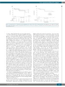
Figure 5. Overall survival (OS) of histological follicular lymphoma (FL) subtypes and FL with and without t(14;18). Kaplan-Meier-plot of the different FL subtypes FL1/2, FL3A, FL3B and diffuse large B-cell lymphoma (DLBCL)/FL3B (A), as well as the comparison of t(14;18)-positive and t(14;18)-negative FL (B) revealed no sig- nificant differences in OS.
Gene expression profiling of FL subtypes
AB
ic events, suggesting that they are biologically distinct.
In order to obtain a more detailed insight into the patho- genesis of FL3A and FL3B, GEP had been performed by Piccaluga et al.8 However, they had not integrated markers for immunohistochemistry and FISH into their study. The main finding of this study was a relatively homogeneous GEP of different FL subtypes. In a supervised analysis approach, Piccaluga et al.8 found that FL1/2 and FL3A formed one cluster, while FL3B formed a separate group distinguishable from FL1/2/3A based on the differential expression of 30 genes. In contrast to these findings, we failed to observe a significant difference in the gene expression patterns of FL1/2 and FL3A on the one hand and of FL3B on the other hand. In contrast, from our data set, a significantly differential gene expression emerged between FL1/2 and FL3A, while FL3B profiles more close- ly resembled those of FL3A. Despite these different find- ings concerning the relationship of FL1/2, FL3A and FL3B, we and Piccaluga et al. identified similar pathways affect- ed in FL1/2 and FL3, mainly targeting cellular metabolism, cell cycle, and cell growth. Applying previously published proliferation signatures14,15 to our FL samples, however, revealed a highly heterogeneous spectrum of proliferation indices in FL1/2 cases in the present study. In keeping with the fact that almost no overlap was observed between the genes from known proliferation signatures and the 643 differentially expressed genes between FL1/2 and FL3A, we provide evidence that proliferation is not the only explanation for the difference between the GEP in FL1/2 and FL3A. An increased proliferation, in particular in FL1/2 samples, had already been described by using miRNA pro- filing, further substantiating the finding of a wide prolifer-
ation spectrum even in FL1/2.22,23
Of 12 genes distinguishing FL1/2 from FL3A/B, all were
over-expressed in FL3A/B. Intriguingly, 3 genes, MRE11A, TXN and TOP2A, had already been associated with the pathogenesis of lymphoma and, therefore, targeting their expression might be beneficial for tailored therapy (Online Supplementary Table S3).24-26
Based on the classification approach performed in the present study, FL1/2 were clearly distinguishable from GCB-DLBCL, while FL3A and FL3B, in contrast, showed
highly similar gene expression patterns; moreover, they both formed an expression cluster intermediate between FL and DLBCL. Piccaluga et al. found that FL3A and FL3B were clearly distinguishable in their gene expression pat- terns in supervised analysis and that both subgroups resembled more closely FL than DLBCL.8 Nevertheless, in their study, they identified 2 FL3B that clustered within the DLBCL group, similar to the findings in the present study.8 These data might indicate that a distinct classifica- tion of FL subtypes based on their GEP is not possible in all cases. Furthermore, these results underline the fact that the grouping of FL3B within FL1-3A might not be biolog- ically justified in all cases. In the Kiel classification system,27 FL3B had been regarded as a follicular variant of DLBCL (“follicular centroblastic lymphoma”).
Since so far only two GEP global studies on FL are avail- able, each one analyzing only a limited number of sam- ples, it is difficult to draw universal conclusions from these investigations and, therefore, additional validation studies are clearly needed. In fact, considering only gene expres- sion data, no clear-cut pattern that might be useful to dis- tinguish tumors with a different follicular component can be obtained. Although only a limited number of FL3B were available to be investigated within the present study, the fact that GEP clearly separated FL1/2 and FL3A/FL3B sug- gests a close relationship between FL3A and FL3B. This notion, however, is in contrast to immunohistochemical and genetic profiles of the different histological FL sub- types that point to a closer relationship between FL1/2 and FL3A, and separating them from FL3B. This phenomenon could possibly be explained by the different methological approaches used, focusing on the examination of tumor cells by immunohistochemistry and FISH, while both tumor and non-malignant bystander cells are simultane- ously interrogated by gene expression profiling.
Finally, the therapeutic implication of a diagnosis of FL3A is still a subject of debate. Many hemato-oncologists regard FL3A as belonging to the spectrum of conventional FL1/2. On the other hand, a recent retrospective analysis of FL3A cases enrolled in the German low- and high-grade lymphoma trials failed to observe any difference in sur- vival between FL3A and FL3B and, most intriguingly,
haematologica | 2018; 103(7)
1189
