
Page 98 - Haematologica June
P. 98
1000
S. Gon et al.
from a large array of genetic and epigenetic alterations in oncogenes and tumor suppressors.5-7 A wealth of informa- tion has emerged from recent pan-(epi)genomic analysis of T-ALLs, and this has allowed mapping of cell-intrinsic genetic defaults in these cells to be expanded. However, much less is known about the potential cell-extrinsic cues that may impact on the leukemia genesis process, includ- ing the role that the TCR might play in malignant trans- formation throughout thymocyte selection, survival and proliferation.8-10 In this study, we sought to address how TCR signaling can interfere or, on the contrary, can be integrated in T-ALL oncogenic networks.
Methods
Patients’ samples
Diagnostic specimens (peripheral blood or bone marrow) col- lected from patients treated at the Timone Children’s Hospital or Paoli Calmettes Institute (Marseille, France) or from Necker Hospital (Paris, France) were used to generate xenografts. Diagnosis and classification were defined by expression of specific
T-cell markers and negativity for B cells and myeloid markers. Healthy human thymus were obtained from Timone Children’s Hospital. Samples were purified by Ficoll-Hypaque centrifugation. T-ALLs were included within FRALLE-2000 or GRAALL-2005 pro- tocols, and informed consent for use of diagnostic specimens for future research was obtained from the patients or relatives in accordance with the Declaration of Helsinki. This study was approved by institutional review boards of all hospitals involved.
Mice
Mice were bred and housed in specific pathogen-free conditions in CIML animal facilities and were handled in accordance with French and European guidelines. Mice strains and oligonucleotides used for mice genotyping are listed in the Online Supplementary Methods and Online Supplementary Table S1. Xenotransplantation of primary human T-ALL samples in immunodeficient NSG mice was performed as previously described.11
TCR-signaling ability assays
TCR-signaling ability assays were performed with 2x10⁷ wild-
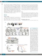
A
B
C
DE
haematologica | 2018; 103(6)
Figure 1. Fit TCRαβ signaling func- tions as a tumor suppressor. (A) Thymus (left) and spleen (right) of typical wild-type (WT) and tumoral Ptendel, [Rag1–/– x Ptendel] and [OT-II x
Rag1–/– x Ptendel] mice. Phenotypes of typical tumors gener- ated by Ptendel in the spleen and by [Rag1–/– x Ptendel] and [OT-II x Rag1–/– x Ptendel] in the thymus. WT and [OT-II x Rag1–/–] controls are shown. CD4 SP or DP gates (top plots, bold squares) were further analyzed for CD3/TCRβ expression (bottom plots). Two typical thymi of [OT-II x Rag1–/– x Ptendel] mice are shown (1 represen- tative of 10). (C) Thymi of indicated mice were analyzed by immunoblot- ting with antibodies specific for Pten, Myc, cleaved Notch1, Bcl2 and Actin as a loading control. #Identification number of analyzed mice. *Mice that did not display T-cell acute lym- phoblastic leukemias (T-ALL) or T-cell lymphoblastic lymphomas (T-LBL) symptoms at the time of sacrifice. (D) Transcriptional downderegulation of transgenic TCRβ chain in [OT-II x Rag1–/– x Ptendel] tumor thymocytes (n=6). Transcripts levels were normal- ized to ABL. Error bars show means with Standard Deviation. Statistical significance was assessed using Mann-Whitney test (**P<0.01). (E) Ptendel mice survival curve was com- pared to [Rag1–/– x Ptendel] or [OT-II x Rag1–/– x Ptendel] mice survival curves using log-rank (Mantle-Cox) test (**P<0.01; ***P<0.001); median weeks of survival are 11 (as previous- ly observed15), 9.95 and 17.9, respec- tively.
(B)
