
Page 50 - Haematologica June
P. 50
M.W. Wlodarski et al.
a DBA silent carrier due to very high erythrocyte adeno- sine deaminase (eADA) levels. The remaining two point mutations c.29T>C; p.Leu10Pro (P5) and c.458A>C; p.Lys153Thr (P6) affect highly conserved residues and, based on results from in silico prediction, are probably deleterious (Online Supplementary Table S1). Biomuta, DMDM, the Exac/GnomAD databases, and NCBI do not report any variants in the codon for Leu10. The GnomAD population database reports one variant in Lys153 (Lys153Arg; rs370700905) identified in 33 out of 232840 total alleles.
Genotype-phenotype association for truncating RPL15 mutations: severe hematologic phenotype and rapid acquisition of treatment independence
All of the individuals with mutations in RPL15 present- ed with typical bone marrow erythroid hypoplasia, ele-
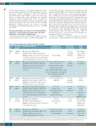
Table 1. Clinical characteristics of patients with RPL15 mutations.
vated eADA, and most of them presented with increased fetal hemoglobin (HbF) levels (Table 1). Notably, hydrops fetalis (considered the most severe hematologic pheno- type of DBA) was associated only with truncating RPL15 mutations. The affected fetuses P2-4 required between four and nine intrauterine transfusions (Table 1). One indi- vidual (P2) with the p.Tyr81* hotspot mutation presented with various physical malformations, while the dysmor- phic features in other patients were less severe (Table 1). Unexpectedly, all 3 patients carrying p.Tyr81* substitution (P1-3) attained a rapid treatment independence both with and without steroid treatment (Figure 2A), while the fourth patient with the RPL15 mutation c.85C>T; p.Gln29* responded to steroids; however, the therapy was discontinued due to overt toxicity.
Based on published observations of genetic revertant mosaicism as a “repair mechanism” in other bone marrow
Pat/ ID (sex)
1/DE071
(F)
2/DE189
(M)
3/DE115
(F)
4/DE202
(M)
5/IL
(F)
6/FR
(M)
RPL15 gene; mutation
c.242dupA;
p.Tyr81*
c.242dupA;
p.Tyr81*
c.242dupA;
p.Tyr81*
c.85C>T;
p.Gln29*
c.29T>C;
p.Leu10Pro
c.458A>C;
p.Lys153Thr
Hematology and therapies
Onset: 3 months old; Hb 4.6g/dL
Lab: MCV↑, eADA↑ (925U/Iec), HbF normal Evolution: spontaneous recovery after 1 transfusion at 6 months old. Relapse at 5.7 years (Hb 3.6g/dL), achieved remission after short course of steroids
Gestational age; malformations; other
37 weeks, IUGR
Age and status at last follow up
6.5 years, normal Hb, no therapies
5 years, normal Hb, no therapies
16 years, normal Hb, no therapies
18 years, regular transfusions
2 years, steroids
22 years, regular transfusions
Family history
Father mutation carrier, eADA↑ (1396U/Iec) but clinically silent
Parents and sister wild type
Parents wild type
Parents and sibling healthy (carrier status unknown), eADA/Hbf unknown Mother mutation carrier: Hb, MCV and HbF normal, eADA unknown
Parents and sibling healthy (carrier status unknown)
Onset: prenatal (4 intrauterine transfusions) Lab: MCV↑, eADA↑ (1526U/Iec), HbF (6.6%) Evolution: 4 transfusions (birth-16 months), achieved remission after short course
34+1 weeks, hydrops fetalis, ptosis, flat nose, deep set ears, intermittent AV-block, duplex left kidney, hypogonadism,
of steroids intersexual genitalia (46XY), microcephaly, left cerebellar hypoplasia, developmental
Onset: prenatal (6 intrauterine transfusions), Lab: MCV↑, eADA↑ (1284U/Iec), HbF↑ (10.6%) Evolution: 2 transfusions after birth, achieved spontaneous remission at age of 4 months Onset: prenatal (9 intrauterine transfusions) Lab: MCV↑, eADA ↑ (2628U/Iec), HbF↑ (2%) Evolution: after birth irregular transfusions, steroid-responsive (4-9 years), discontinued due to toxicity, transfusion-dependent from age of 9 years
Onset: 4 months old, Hb 5g/dL
Lab: MCV↑, eADA and HbF unknown
Evolution: transfusion dependent, recently started steroids with good response
Onset: 6 months old, Hb 7g/dL
Lab: MCV normal, eADA unknown, HbF↑ (15%) Evolution: initially steroid-responsive, transfusion dependent from age of 5 years
disorder with mental retardation
and cerebral palsy
35+6 weeks, hydrops fetalis, IUGR, PFO
32+3 weeks, hydrops fetalis, hypogammaglobulinemia, plantar warts during steroid therapy
27 weeks (placenta previa), none
41 weeks, low-set hair line; growth retardation and mental retardation
952
All patients presented with DBA-typical erythroblastopenia in bone marrow. Pat: patient number; ID: patient identifier in respective national registry; RP: ribosomal protein; F: female, M: male; Hb: hemoglobin; Lab: supportive laboratory parameters; MCV: mean corpuscular volume; eADA: erythrocyte adenosine deaminase; Evolution: evolution of dis- ease and therapies; ↑ elevated for age; HbF: fetal hemoglobin; Remission: treatment independence; IUGR: intrauterine growth restriction; PFO: persistent foramen ovale.
haematologica | 2018; 103(6)
