
Page 167 - Haematologica June
P. 167
Therapeutic in vivo assays in mouse xenografts
OCI-Ly7, L363 and UPN1 tumor cells (5x10⁶ cells per animal) were injected subcutaneously in 6-week-old male immunodefi- cient Rag2-/-IL2gc-/- mice. Eight animals per group were used on each experiment. Once tumors reached volumes of 100 mm3, treat- ment was started by intratumorally injecting 50, 200 or 400 mg of the peptide (a truncated peptide was used for control mice at the same dosage). Tumor size was measured every 2 days, as described previously.47 When tumors reached volumes of 2000
mm3, animals were euthanized.
Statistical Analysis
Results are expressed as mean ± SEM. At least three independ- ent experiments for cell viability, apoptosis and pHi assessment assays were performed (in duplicate each). The normal distribu- tion of values was assessed by using the Shapiro-Wilks and Kolmogorof-Smirnov tests, and the statistical significance of differ- ences was determined with the student’s-t test, taking two-tailed P<0.05 as the criterion for significance. For in vivo experiments, data were analyzed by two way ANOVA test and the Bonferroni post-tests to compare replicate means. Statistical analyses were performed using the Graph Pad Prism 5 program.
Results and Discussion
Generation and characterization of functional peptides targeting AE2
A series of 24 linear peptides were designed to bind a short, highly conserved region in human and mouse AE2 (Figure 1A). Once synthesized, peptides were tested for their ability to inhibit the suppressor activity of natural CD4+CD25+Treg cells in vitro. Thus, CD4+CD25– effector T cells activated with anti-CD3 monoclonal antibody in the presence of Treg cells were used to analyze the capacity of each peptide to restore T-cell proliferation inhibited by Treg cells. Among the 24 peptides, three (p17AE2, p19AE2 and p20AE2) were able to restore and even enhance the prolif- eration of effector T cells (Figure 1B). Surface plasmon res-
Peptides specifically targeting AE2 induce apoptosis in tumor
onance showed a dose-dependent binding of the p17AE2 peptide to a 36 amino acids long peptide encompassing the third extracellular loop of human AE2 that is crucial for its exchange function,25,34 thus revealing physical binding (Figure 1C). The p17AE2 peptide was therefore selected for further experiments.
Functional targeting peptide p17AE2 shows opposite functions in different T-cell subsets
Next, we tested the effect of p17AE2 on conventional effector T-cell proliferation in the absence of Treg cells. In vitro experiments showed that the addition of p17AE2 to murine effector T cells slightly increased cell prolifera- tion in response to anti-CD3 stimulation, and similarly promoted IL-2 secretion, while apoptosis was not affect- ed (Figures 2A). This capacity to promote effector T-cell proliferation was also observed in human effector T cells from healthy donors stimulated with anti-CD3/CD28 beads (Figure 2B). Conversely, p17AE2 reduced cell prolif- eration of activated Treg cells in culture and induced cell apoptosis, while the production of IL-10 was not signifi- cantly altered, (Figures 2C). Likewise, p17AE2 decreased cell viability of the human-derived T-cell leukemia cell line Karpas299, which shows characteristics typical of natural Treg cells with a CD4+CD25+Foxp3+ phenotype.48 On the other hand, p17AE2 did not affect cell survival of Jurkat T-cell leukemia cells with a CD4+CD25– T-cell effector phenotype (Figure 2D).39 We then determined whether changes in cell survival were related to varia- tions of the pHi. Upon incubation with p17AE2, the pHi in effector T lymphocytes remained similar to that in con- trol cells or cells treated with the truncated peptide, while pHi values decreased in Treg cells over time (Figure 2E).
AE2 targeting promotes apoptosis of B-cell leukemia, lymphoma and multiple myeloma cells
The effect of the p17AE2 peptide in vitro was also eval- uated on peripheral blood B lymphocytes isolated from healthy donors as well as on cell lines derived from
ABC
D
E
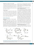
Figure 4. Modulation of the exchanger function of AE2 driven by p17AE2 peptide. (A) Basal pHi in human peripheral blood B lymphocytes (PBLs) and tumor B-cell lines. (B) Changes in pHi values upon treatment with p17AE2. (C) Average basal pHi in sensitive and resistant tumor B-cell lines. (D-E) Effect of p17AE2 treatment on the AE2 activity in sensitive Jeko1 and resistant U266 cell lines. ***P<0.001.
haematologica | 2018; 103(6)
1069
