
Page 166 - Haematologica June
P. 166
J. Celay et al.
In vitro assays for murine effector and regulatory T cells Regulatory (CD4+CD25+) and effector (CD4+CD25−) T cells were isolated from BALB/c mouse spleens using Treg isolation kits (Miltenyi Biotec). For the screening of the peptides targeting AE2, CD3-stimulated effector T cells were cultured in the presence or absence of purified Treg cells and peptides, as previously described.43 Then, cell proliferation was calculated by adding [methyl-3H] thymidine, and incorporated radioactivity was meas- ured using a scintillation counter.44 The same procedure was car- ried out to determine the effect of p17AE2 in effector and regula- tory T cell populations. IL-2 and IL-10 secreted to the supernatant
of cell cultures were measured by ELISA (Pharmingen).
Intracellular pH measurement
To asses pHi, cells were stained with the intracellular fluorescent pH indicator BCECF-AM (Biotium), as described.32,45 Briefly, cells were stained and washed in MACS buffer supplemented with the corresponding peptide at a final concentration of 50 mg/mL. Then, cells were excited at 488 nm and the ratio of emission wavelengths 530/661 nm was determined in a FACS Calibur cytometer. The
AB
nigericin clamp technique was used to estimate pHi values from calibration curves as described.31,46
Determination of anion exchange activity
To determine the Cl−/HCO − exchange activity, cells were 3
stained with BCECF-AM in Krebs-Ringer bicarbonate buffer (KRB, which contains in mM: 115 NaCl, 4.7 KCl, 1.5 CaCl2, 25 NaHCO3, 1 Na+-pyruvate, 1.2 KH PO ,1 MgSO , 5 glucose, and
244
carbogen − 95% CO2 + 5% O2) and incubated as described
above. Then, cells were washed and resuspended in Cl–-free buffer
(in mM: 115 Na+-isethionate, 4.7 K+-gluconate, 1.5 Ca2+-gluconate,
25 NaHCO , 1 Na+-pyruvate, 1.2 KH PO , 1 MgSO , 5 glucose, 3244
and carbogen) supplemented with 50 mg/mL of the corresponding
peptide, and pHi was measured every 2 minutes by flow cytome-
try. Cl– was replaced by isothionate in order to maintain osmolali-
ty. Changes in anion exchange activity were represented as the
variation of pH along the time after extracellular Cl– removal. This
i
––
removal forces the efflux of Cl and the influx of HCO , and intra-
3
cellular pH increases as a result of a reverse activity of the AE2
anion exchanger.29,32
C
E
D
F
GH
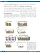
Figure 3. Effect of p17AE2 on human normal B lymphocytes and tumor B-cell lines. (A) Cell viability of CD19+ cell isolated from human peripheral blood lymphocytes (PBLs) upon treatment with 5, 10 or 50 mg/mL of truncat- ed or p17AE2 peptides. (B) Effect of p17AE2 on cell viability of human B-cell leukemia, lym- phoma and multiple myeloma cell lines. Cell viability, apoptosis, and cell cycle abnormali- ties in tumor B-cell (C, D, E), AML (F) and T-ALL cell lines (G, H) after 48 hour incubation with p17AE2 peptide. *P<0.05; **P<0.01; ***P<0.001.
1068
haematologica | 2018; 103(6)
