
Page 129 - Haematologica June
P. 129
MYD88L265P mutation detection by ddPCR on BM, PB and ctDNA
IgM-LPL, 10 IgG-LPL, 4 IgM-MGUS): 194 samples were taken at baseline in active disease (128 BM, 66 PB) and 97 samples during follow up (43 BM and 54 PB). The baseline samples were defined either “treatment-naïve” (143/194, 73.7%, 99 BM and 44 PB) or “relapsed” (51/194, 26.2%, 29 BM and 22 PB), according to previous exposure to chemo- immunotherapy. All the follow-up samples were taken for the purpose of studying MRD at different time points after specific treatment. The distribution of the study pop- ulation is summarized in Online Supplementary Figure S1, while the patients’ main clinical features are detailed in Table 1.
Detection limit of droplet digital polymerase chain reaction
The detection limit of ddPCR was determined using a serial dilution of MYD88L265P mutated gDNA in WT DNA at levels of 35, 3.5, 0.35, 0.035 and 0.0035%, correspon- ding to 10500, 1050, 105, 10.5 and 1 mutated copy pres- ent in 100 ng of gDNA (30,000 copies) which is the over- all quantity of gDNA loaded per well. We identified the limit of detection using a statistical method based on binomial distribution, as previously reported.29 This analysis indicated that we were able to detect the muta- tion, with a good degree of confidence, when the level of mutated copies was more than 0.035% (10 mutated copies in 30000 WT). This value mirrored the cut-off mutated:WT ratio we identified based on the control group (40 healthy subjects) (Figure 1A). Additional repro- ducibility tests confirmed that the above calculated limit of detection and the experimentally detected cut-off, emerging from the control group, are equally reliable (Online Supplementary Table S2).
MYD88L265P screening by droplet digital polymerase chain reaction in baseline samples
Overall 142/148 (96%) patients were identified as hav- ing the MYD88L265P mutation. We observed a 91.6% muta- tion rate (33/36) among relapsed patients and 97.3% rate (109/112) among treatment-naïve patients (P=0.15). Notably, no one of the 6 MYD88L265P WT patients (4 WM with histological BM invasion of 20%, 30%, 30% and 60%) or 2 patients with IgG LPL (with 20% and 30% his- tological BM invasion) showed either alternative MYD88 or CXCR4 mutations, as investigated by Sanger sequenc- ing on unselected cells.10,12,30 All BM samples from 20 patients with multiple myeloma used as negative controls were below the limit of detection (defined as previously described) and below the mutation cut-off ratio estab- lished based on 40 PB samples from healthy individuals (mutated:WT ratio <3.4x10⁻⁴) (Figure 1A). Moreover, to confirm the specificity of our assay for mutational screen- ing, 15 patients with mantle cell lymphoma, 10 with fol- licular lymphoma and 10 with chronic lymphocytic leukemia were tested for the MYD88L265P mutation. All samples from the patients with follicular lymphoma or mantle cell lymphoma were WT, whereas one of the 10 patients with chronic lymphocytic leukemia showed a mutation ratio of 4.4x10⁻⁴, as already described in this dis- ease.6
Looking at the single tissues, 95.3% (122/128) of base- line BM and 71.2% (47/66) of PB samples scored positive for MYD88L265P (BM median mutation burden 3.60x10⁻², range: 2.00x10⁻⁴ – 7.30 x10⁻¹; PB median 5.00x10⁻³, range: 1.00x10⁻⁴ – 2.80 x10⁻¹) (Figure 1A). Notably, among the PB
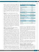
Table 1. Patients’ clinical and biological features at baseline. Patients’ chacteristics
Baseline samples (n= 148)
Waldenström macroglobulinemia
IgG-lymphoplasmacytic lymphoma IgM-MGUS
IgM-LPL amyloid tumor
Sex, female
Median age (years, range) Median hemoglobin (range), g/dL
Median IgM (range), g/dL
133 (89.8%)
10 (6.7%)
4 (2.8%)
1 (0.7%) 51 (34%) 67 (24-88) 11.5 (7.5-16.8)
Waldenström macroglobulinemia
MGUS
2.397 (0.233-12.5)
0.785 (0.492-2.252)
Median IgG (range), g/dL
IgG-lymphoplasmacytic lymphoma 1.958 (0.916-3.447) Median β2 microglobulin (range), mg/L 2.62 (1-8.83)
Bone marrow biopsy
Bone marrow flow cytometry
Splenomegaly
Adenopathies
Organomegaly
40% (0-95%)
12% (0-87%)
18 (12%)
29 (19.5%)
Median bone marrow infiltration
haematologica | 2018; 103(6)
LPL: lymphoplasmacytic lymphoma; MGUS: monoclonal gammopathy of undeter- mined significance.
samples there was a statistically significant difference in mutation rate between samples from relapsed patients (median burden 4.00x10⁻⁴, range: 1.00x10⁻⁴ – 1.00x10⁻³) and treatment-naïve patents (median burden 2.80x10⁻³, range: 2.00x10⁻⁴ – 1.00x10⁻²), supporting the possible negative impact on mutation detection of previous treatment on PB samples (P<0.0001) (Figure 1B).
Seventy-four patients in this series had paired baseline BM and PB samples. Overall, in this subgroup of patients, the rate of MYD88L265P mutation detection by ddPCR was 93% (69/74) on BM and 72% (53/74) on PB samples. Accordingly, the detection rates were higher among treat- ment-naïve patients than among relapsed patients: 95% (52/55) on BM and 82% (45/55) on PB versus 89% (17/19) on BM and 42% (8/19) on PB, respectively (P=0.014). In addition, 15 of these 74 patients (20%) showed BM- mutated/PB-WT discordance, such discordance being more frequent among relapsed cases than among treat- ment-naïve cases (8/19, 42% vs. 7/55, 13%; P=0.02) (Figure 1C).
Finally, a test of within-run reproducibility showed uni- form results across the experiments, with an inter-assay Standard Deviation (SD) of 1.3% for the MYD88L265P posi- tive control and 0.01% for WT gDNA samples.
Comparison of MYD88L265P droplet digital polymerase chain reaction versus allele-specific quantitative polymerase chain reaction assays
Once the ddPCR assay had been optimized, the sensi- tivity of the MYD88L265P ddPCR was compared to that of ASqPCR on a standard curve of 10-fold serial dilutions constructed with a highly MYD88L265P mutated WM sam- ple (70%, mutated/WT ratio 7.00x10⁻¹, diluted to 35%),
1031
