
Page 103 - Haematologica June
P. 103
αβTCR signaling acts as a tumor suppressor
and developed T-ALL, acquired a deficiency in TCR sig- naling. Unstimulated primary mouse T-ALLs tend to quickly undergo apoptosis in liquid culture in vitro and can be rescued through anti-CD3/anti-CD28 TCR activation, as shown for the TCRαβ+ Cdkn2a–/– T-ALL mouse model (Figure 4A, Online Supplementary Table S6 and Online Supplementary Figures S5 and S6A). By contrast, no rescue could be obtained for T-ALL blasts from the Ptendel T-ALL model, and TCRαβ+ Ptendel leukemic cells quickly died with or without stimulation. To further investigate the impact of TCR triggering at the molecular level, freshly harvested tumors were CD3/CD28 stimulated for 2 min and analyzed by immunoblotting with antibodies specific for phosphorylated tyrosine species (P-Tyr) (Figure 4B). In control purified DP and non-purified (NP) mouse WT thy- mocytes, activation of the TCR pathway triggered various intracellular signaling molecules23 leading to a marked increase in the pattern of global tyrosine phosphorylation (Figure 4B and C), as previously described.24 Similar results were obtained with Cdkn2a–/– T-ALLs, in line with prolif- eration data described above. By contrast, global tyrosine phosphorylation was significantly dampened in murine TCRαβ+ Ptendel T-ALLs (Figure 4B and C). However, P-Tyr antibody does not detect the activation of AKT which is the main downstream target of Pten.12 Thus, we specifical- ly monitored phosphorylation of Akt at Ser473. It was previously showed that Akt phosphorylation was very high in non-tumoral Pten-deficient thymocytes compared to Pten-proficient thymocytes.13 However, we found that P-Akt levels in Ptendel T-ALL are similar to the one detected for WT thymocytes and Cdkn2a–/– T-ALL (Figure 4B and C) and thus are lower than one might expect from Pten- deficient thymocytes.13
It is noteworthy that when we induced inactivation of Pten in Cdkn2a–/– T-ALL cells, the ability of those cells to proliferate upon stimulation was conserved (Online Supplementary Figure S7), suggesting that once pre-tumoral thymocytes have passed selection and the tumor is estab- lished, late deletion of Pten no longer interferes with TCR- mediated activation. In the same line, T cells from disease- free Ptendel spleen were able to proliferate upon anti- CD3/28 stimulation (Online Supplementary Figure S8), con- firming that, per se, Pten loss is not directly responsible for the TCR signaling inhibition observed in Ptendel T-ALL. Together with the fact that human late cortical T-ALLs are frequently carrying PTEN loss-of-function alterations,5 this pointed to a possible role of Pten loss in the dysregu- lation of the selection process. We thus assessed whether the above observation indicating that TCR signaling is impaired in Pten-deficient T-ALL was relevant in human primary T-ALL samples. To obtain adequate quantities of viable human leukemia cells devoid of contaminating residual physiological mature T cells, samples from T-ALL patients were engrafted into immunodeficient NSG mice. In our patient-derived xenograft (PDX) collection, PTEN was present in all TCRneg T-ALL, while it was not expressed in 5 out of 6 TCRαβ+ samples. We investigated the 5 PTEN-deficient TCRαβ+ T-ALL (T-ALL 8, 9, 35, 38 and 47; their corresponding xenografts were denoted Xg8, Xg9, Xg35, Xg38 and Xg47), and 5 TCRneg T-ALLs were used as controls (Xg3, Xg13, Xg20, Xg23 and Xg40) (Online Supplementary Table S7 and Online Supplementary Figure S6B). Leukemic grafts were harvested from mice and stimulated with anti-CD3 and anti-CD28. In contrast to mouse, human T-ALL do not die quickly in liquid cul-
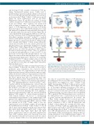
Figure 5. Model for integration of Pten loss-of-function and TCR signaling-medi- ated tumor suppression. In the context of Pten loss, thymocytes bearing fit or high affinity TCR would be eliminated while those bearing no/low affinity TCR would be rescued from death-by-neglect. However, harboring TCR complex that does not signal properly would prevent further thymocyte differentiation, provid- ing an additional opportunity for malignant transformation.
ture; thus we assessed the impact of TCR stimulation at the cellular level. We observed that activation-induced cell death (AICD) was triggered for only 1 TCRαβ+ T-ALL (Xg35), the remaining 4 TCRαβ+ T-ALL, as well as control TCRneg T-ALLs, being resistant to AICD (Figure 4D and E). To investigate signaling downstream of the TCR, as described above for mouse T-ALL, PDX cells were lysed 2 min post activation with anti-CD3/CD28, and analyzed by immunoblotting. In control (disease-free) non-purified (NP) or purified CD4 SP human thymocytes, activation of the TCR pathway led to a marked increase in the pattern of global tyrosine phosphorylation. In contrast, the TCRαβ+ T-ALLs samples showed a reduced and some- what intermediate activation of tyrosine phosphorylated species compared to NP or CD4 SP thymocytes and TCRαβneg controls (Figure 4F). Globally, the P-Tyr activa- tion index of TCRαβ+ T-ALL was significantly lower than physiological controls (NP or CD4 SP; P<0.05) but not sig- nificantly higher than TCRαβneg T-ALL (Figure 4G). In con- trast, AKT phosphorylation was detected in PTEN-defi- cient TCRαβ+ T-ALLs in unstimulated conditions, then fol- lowing stimulation, it was slightly increased (approx. 3.5- fold) and ended up equivalent to CD4 SP positive control (Figure 4G).
haematologica | 2018; 103(6)
1005
