
Page 43 - Haematologica Vol. 107 - September 2022
P. 43
REVIEW ARTICLE - IgM monoclonal gammopathies of clinical significance J. Khwaja et al.
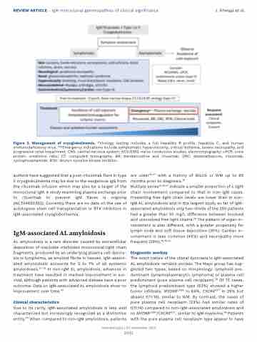
Figure 2. Management of cryoglobulinemia. *Virology testing includes a full hepatitis B profile, hepatitis C, and human immunodeficiency virus. **Emergency indications include symptomatic hyperviscosity, critical ischemia, severe neuropathy, and progressive renal impairment. CNS: central nervous system; NCS/EMG: nerve conduction studies, electromyography; uPCR, urine protein creatinine ratio; CT: computed tomography; BR: bendamustine and rituximab; DRC: dexamethasone, rituximab, cyclophosphamide; BTKi: Bruton tyrosine kinase inhibitor.
authors have suggested that a post-rituximab flare in type II cryoglobulinemia may be due to the exogenous IgG from the rituximab infusion which may also be a target of the monoclonal IgM. A study examining plasma exchange prior to rituximab to prevent IgM flares is ongoing (NCT04692363). Currently there are no data on the use of autologous stem cell transplantation or BTK inhibitors in IgM-associated cryoglobulinemia.
IgM-associated AL amyloidosis
AL amyloidosis is a rare disorder caused by extracellular deposition of insoluble misfolded monoclonal light chain fragments, produced by an underlying plasma cell dyscra- sia or lymphoma, as amyloid fibrils in tissues. IgM-associ- ated amyloidosis accounts for 5 to 7% of all systemic amyloidoses.35-39 In non-IgM AL amyloidosis, advances in treatment have resulted in marked improvement in sur- vival, although patients with advanced disease have a poor outcome. Data on IgM-associated AL amyloidosis show no improvement over time.40
Clinical characteristics
Due to its rarity, IgM-associated amyloidosis is less well characterized but increasingly recognized as a distinctive entity.40 When compared to non-IgM amyloidosis, patients
are older36,37 with a history of MGUS or WM up to 65 months prior to diagnosis.38
Multiple series36,38,41 indicate a smaller proportion of λ light chain involvement compared to that in non-IgM cases. Presenting free light chain levels are lower than in non- IgM AL amyloidosis and in the largest study so far of IgM- associated amyloidosis only two-thirds of the 250 patients had a greater than 50 mg/L difference between involved and uninvolved free light chains.40 The pattern of organ in- volvement is also different, with a greater propensity for lymph node and soft tissue deposition (35%). Cardiac in- volvement is less common (45%) and neuropathy more frequent (28%).36,38,40
Diagnostic workup
The exact nature of the clonal dyscrasia in IgM-associated AL amyloidosis remains unclear. The Mayo group has sug- gested two types, based on morphology; lymphoid pre- dominant (lymphoplasmacytic lymphoma) or plasma cell predominant (pure plasma cell neoplasm).38 Of 75 cases, the lymphoid predominant type (63%) showed a higher tumor infiltrate, MYD88L265P in 84%, CXCR4MUT in 29% but absent t(11;14), similar to WM. By contrast, the cases of pure plasma cell neoplasm (23%) had similar rates of t(11;14) compared to non-IgM-associated amyloidosis and no MYD88L265P/CXCR4MUT, similar to IgM myeloma.38 Patients with the pure plasma cell neoplasm type appear to have
Haematologica | 107 September 2022
2042
