
Page 190 - Haematologica Vol. 107 - September 2022
P. 190
ARTICLE - NOTCH2 in myeloma-derived extracellular vesicles D. Giannandrea et al.
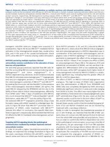
Figure 4. Molecular effects of NOTCH2 modulation on multiple myeloma cell-released extracellular vesicles. (A) Western blot analysis confirms the knockdown (KD) efficacy on NOTCH2-TM levels in human multiple myleoma cell lines (HMCL) and the pro- duced multiple myeloma extracellular vesicles (MM-EV). a-tubulin and TSG101 were used as loading controls for cell and vesicle protein extracts, respectively. (B) Nanoparticle tracking analysis (NTA) on MM-EVSCR and MM-EVN2KD from HMCL does not show significant changes in concentration and size; D50=size point below which 50% of the extracellular vesicles (EV) are contained. Data are expressed as mean value ± standard error of the mean of at least 4 experiments (RPMI8226 n=4; OPM2 n=6). Statistics by two-tailed t-test did not show any significant difference. (C) MM-derived EV activate NOTCH signaling in receiving cells: a NOTCH reporter assay was carried out on HeLa cells stimulated with MM-EVSCR and MM-EVN2KD from HMCL (EV derived from OPM2 cells), or control fresh medium (w/o EV). Luciferase activity is expressed as the ratio between Nano/Firefly luciferase lumi- nescence units. Data are expressed as mean value ± standard error of 4 experiments. Statistics by ANOVA and Tukey post-test: *P<0.05. (D) Activation of NOTCH signaling in the trunk of Tg(T2KTp1bglob:hmgb1-mCherry) zebrafish embryos (zf) 4 hours after the injection of MM-EVSCR and MM-EVN2KD (EV derived from RPMI8226 cells), or control fresh medium (w/o EV). Representative pictures of each condition are reported on the left (20x and 60x magnification, the upper and the lower respectively); a graph on the right represents the mean value +/- standard error of the mean of the corrected total fluorescence (CTF) measured in caudal hematopoietic tissue (CHT). In particular four in vivo experiments involved zf embryos injected with negative control (n=20) or MM-EVSCR (n=27) and MM-EVN2KD (n=27). Statistics by ANOVA and Tukey post-test excluding outliers identified through the ROUT method (Q=1%): *** P<0.001.
transgenic zebrafish embryos. Images were acquired 4 h postinjection. Figure 4D shows MM-EVSCR mediated NOTCH activation in the intersegmental vessels (Se), caudal artery (CA), and in the area of the caudal hematopoietic tissue (CHT), while MM-EVN2KD induces a barely visible stimulation.
NOTCH2 carried by multiple myeloma-derived extracellular vesicles contributes to the education of bone marrow cell populations
We and other groups previously reported that MM cells af- fect the surrounding BM microenvironment inducing osteo- clastogenesis22-24 and tumor angiogenesis16,21 in a NOTCH-dependent way. Moreover, recent evidence indicates that MM-EV stimulate osteoclastogenesis,13,31-33 angiogenesis and carry pro-angiogenic proteins.11,34 Therefore, we verified the osteoclastogenic potential of MM-EVSCR and MM-EVN2KD by treating the monocyte cell line Raw264.7 in the presence of the osteoclastogenic chemokine RANKL (30 ng/mL). We used MM-EV released by the RPMI8226 cell line due to the ability of these cells to induce osteoclastogenesis, differently from OPM2 cells.22 After 7 days of treatment with MM-EV, OCL count showed that MM-EVSCR induced Raw264.7 cell dif- ferentiation, while MM-EVN2KD lost this ability (Figure 5A). We assessed MM-EVN2KD angiogenic potential using a tube formation assay with HPAEC seeded on a Matrigel layer. Re- sults in Figure 5B show that MM-EVSCR promotes tube or- ganization of HPAEC, while treatment with MM-EVN2KD reduces this effect. In conclusion, this specific RNA inter- ference approach unequivocally demonstrated that NOTCH2 KD affects the MM-EV mediated osteoclastogenesis and an- giogenesis.
Targeting NOTCH signaling blocks the pathological communication mediated by multiple myeloma-derived extracellular vesicles
In order to provide a higher translational potential to our findings, we exploited the strategy illustrated in Figure 6A. We used γ-secretase inhibitors (50 μM DAPT), used in re- search works and clinics to inhibit pan-Notch signaling15 to
block NOTCH activation in EC and OCL induced by MM-EV. Figure 6B and C clearly show that MM-EV induce angiogen- esis and osteoclastogenesis in a NOTCH-dependent way. In consideration of the well known osteoclastogenic and an- giogenic roles of NOTCH signaling,16,21-24 we planned our ex- periments to distinguish the effect of the endogenous and vesicular NOTCH. Indeed, if we compare the effect of DAPT on osteoclastogenesis (Figure 6B) in the absence of EV and suboptimal concentration of RANKL, we can see that it ab- rogates OCL differentiation with a non-statistically signifi- cant decrease, while DAPT abrogates much higher levels of osteoclastogenesis induced by MM-EV (+250%) in a statis- tically significant way, indicating that the greater effect of MM-EV is NOTCH dependent.
The effect of DAPT on MM-EV-induced angiogenesis was analogous. The high increase of HPAEC tube organization upon the administration of MM-EV from RPMI8226 and OPM2 cells was completely abrogated by DAPT, showing a statistically significant reduction in areas and nodes (ranging from 29,5% to 51,3%). This effect was clearly higher than the slight inhibitory trend observed on basal angiogenesis upon DAPT administration (Figure 6C). We also ruled out that the obtained results could be due to the toxic effect of DAPT on OCL and EC (Online Supplementary Figure S6).
Overall, these results confirm that the studied biological ef- fects of EV are NOTCH mediated and can be blocked by treatment with γ-secretase inhibitors.
Finally, in order to strengthen the translational potential of our in vitro findings, we reasoned that the BM of MM pa- tients does not contain only MM-derived EV, but EV derived from the whole MM-educated BM cell populations. There- fore, in order to confirm the NOTCH-dependent role of EV in the pathological communication occurring in the BM of MM patients, we got advantage of EV from BM aspirates of patients with the benign MGUS (MGUS-BM-EV) or MM (MM-BM-EV) (Online Supplementary Table S1), which may recapitulate the complexity of the BM microenvironment. We compared the angiogenic potential of HPAEC un- treated or treated with MGUS-BM-EV or MM-BM-EV. Fig-
Haematologica | 107 September 2022
2189
