
Page 186 - Haematologica Vol. 107 - September 2022
P. 186
ARTICLE - NOTCH2 in myeloma-derived extracellular vesicles D. Giannandrea et al.
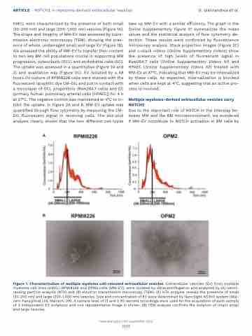
HMCL were characterized by the presence of both small (30-200 nm) and large (200-1,000 nm) vesicles (Figure 1A). The shape and integrity of MM-EV was assessed by trans- mission electronic microscopy (TEM), showing the pres- ence of whole, undamaged small and large EV (Figure 1B). We assessed the ability of MM-EV to transfer their content to two key BM cell populations crucial in supporting MM progression, osteoclasts (OCL) and endothelial cells (EC). The uptake was assessed in a quantitative (Figure 2A and 2) and qualitative way (Figure 2C). EV isolated by a 48 hours (h) culture of RPMI8226 cells were stained with the fluorescent lipophilic dye CM-DIL and put in contact with a monolayer of OCL progenitors (Raw264.7 cells) and EC (primary human pulmonary arterial cells [HPAEC]) for 4 h at 37°C. The negative control was maintained at 4°C to in- hibit the uptake. In Figure 2A and B, MM-EV uptake was quantified through flow cytometry by measuring the CM- DIL fluorescent signal in receiving cells. The dot plot analysis clearly shows that the two different cell types
take up MM-EV with a similar efficiency. The graph in the Online Supplementary Figure S1 summarizes the mean values and the statistical analysis of flow cytometry de- tection. These results were confirmed by fluorescence microscopy analysis. Stack projection images (Figure 2C) and z-stack videos (Online Supplementary Videos) show the presence of high levels of fluorescent signal in Raw264.7 cells (Online Supplementary Videos S1) and HPAEC (Online Supplementary Videos S2) treated with MM-EV at 37°C, indicating that MM-EV may be internalized by these cells. As expected, internalization is blocked when cells are kept at 4°C, suggesting that an active pro- cess is involved.
Multiple myeloma-derived extracellular vesicles carry NOTCH2
Due to the important role of NOTCH in the interplay be- tween MM and the BM microenvironment, we wondered if MM-EV contribute to NOTCH activation in BM cells by
A
B
Figure 1. Characterization of multiple myeloma cell-released extracellular vesicles. Extracellular vesicles (EV) from multiple myeloma cell lines (HMCL) RPMI8226 and OPM2 cells (MM-EV), were isolated by ultracentrifugation and analyzed by (A) nanot- racking particle analysis (NTA) and (B) electron transmission microscopy (TEM). (A) NTA analysis reveals the presence of small (30-200 nm) and large (200-1,000 nm) vesicles. Size and concentration of EV were determined by NanoSight NS300 system (Mal- vern Panalytical Ltd, Malvern, UK). A camera level of 12 and 5 30-second recordings were used for the acquisition of each sample of 3 independent EV isolations and one representative image is shown. (B) TEM analysis confirms the isolation of intact small and large vesicles.
Haematologica | 107 September 2022
2185
