
Page 199 - 2022_02-Haematologica-web
P. 199
LETTERS TO THE EDITOR
SARS-CoV-2 infection in aplastic anemia
Coronavirus disease 2019 (COVID-19), caused by severe acute respiratory syndrome coronavirus-2 (SARS- CoV-2), was declared a pandemic by the World Health Organization in March 2020. Compared to patients with non-hematologic cancers, patients affected by hemato- logic disorders have increased mortality and more pro- longed viral RNA persistence.1-3 Since the early phase of the pandemic, several groups have described thrombocy- topenia or secondary hemophagocytic lymphohistiocyto- sis in patients infected by SARS-CoV-2, problems likely due to a cytokine storm and the potential cytotoxicity of the virus.4,5
Aplastic anemia (AA), a rare autoimmune disease with an incidence of two cases per million population, is char- acterized by cytopenia and bone marrow hypocellularity.6 It has been proposed that, in acquired AA, an initiating event provokes an aberrant immune response, triggering oligoclonal expansion of cytotoxic T cells that destroy hematopoietic stem cells.
The consequences of SARS-CoV-2 infection in known cases of AA are not clear.7 Additionally, it is unknown whether this virus can trigger an aberrant immune response leading to depletion of the stem cell compart- ment and inducing bone marrow failure. Here we describe the features and clinical outcome of a group of patients affected with AA and SARS-CoV-2 infection between April 2020 and January 2021.
A national survey was launched in April 2020 to assess the clinical features and outcome of patients with pre- existing AA and new onset AA after SARS-CoV-2 infec- tion. The criteria for diagnosing AA and classifying its severity were described previously by Camitta et al.8 The diagnosis of SARS-CoV-2 infection was confirmed by nasopharyngeal swab9 at the onset of symptoms or at access to the hematology department.
The study population consisted of 23 patients with AA (30% with very severe AA, 26% with severe AA and 43% with non-severe AA) with a median age of 49 years (range, 20–77); there were seven females and 16 males. All cases were acquired, except one with Fanconi anemia. A subclinical paroxysmal nocturnal hemoglobinuria clone was present in five cases. None of the patients was vaccinated against SARS-CoV-2.
At the onset of SARS-CoV-2 infection, 60% (14/23) of the patients were on active immunosuppressive therapy – six on high-dose cyclosporine maintenance treatment after horse antithymocyte globulin, one on eltrombopag and cyclosporine and seven on a combination of cyclosporine and mycophenolate mofetil – as part of graft-versus-host disease prophylaxis after a reduced- intensity allograft. Table 1 summarizes the populations' demographic and allogeneic stem cell transplant details.
The most common symptoms were fatigue, general malaise, fever, dry cough, shortness of breath, loss of smell, and diarrhea; 29% (7/23) of patients who devel- oped a COVID-19-defining event (6 pneumonia and 1 hepatitis) were hospitalized (median 5 days; range, 3-12). Within this subgroup, three patients required oxygen supplementation, of whom two needed escalation to intensive care unit admission for high-flow oxygen and monitoring, but none required mechanical ventilation.
At diagnosis of the infection, median blood count parameters showed pancytopenia: white blood cells 2.3x10⁹/L (range, 0.42–5.1; interquartile range [IQR], 0.97), neutrophils 1.08x10⁹/L (range, 0.14–2.56; IQR, 0.68), hemoglobin 93.5 g/L (range, 74–139; 25th per-
centile 82, 75th percentile 101) and platelets) 35x109/L (range, 2-121; IQR, 42). None developed evidence of sec- ondary hemophagocytic lymphohistiocytosis.
Upon review of blood results prior to the SARS-CoV-2 infection, it was possible to appreciate a progressive decline in all hematologic indices consistent with overt relapse (confirmed by bone marrow hypocellularity meeting diagnostic criteria) in two patients and, although not meeting relapse criteria, requiring treatment, intense monitoring, and transfusion support in 15 patients.
Interestingly, three cases (12.5%) of idiopathic AA were diagnosed a few weeks after documented SARS- CoV-2 infection. Blood counts performed in the immedi- ate past for other medical reasons showed normal para meters in all these three patients, who developed severe or very severe AA with heavy transfusion dependency, and eventually required treatment with immunosuppres- sive therapy or hematopoietic stem cell transplantation (Table 1). At the time of reporting, all three patients are in remission with good hematologic response.
Figure 1 shows the median values of white blood cell
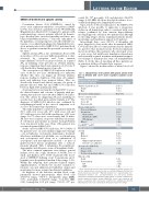
Table 1. Characteristics of the patients with aplastic anemia at the time of infection with severe acute respiratory syndrome coron- avirus-2.
AA disease characteristics at SARS-Cov-2 infection
Number of patients (n, %) Female
Male
Age in years, median [range]
Disease category, n (%) Very severe AA Severe AA Non-severe AA
Disease status (n, %) New onset/diagnosis In remission
On treatment
On CSA after hATG Eltrompopag
Others* Post-HSCT** on IST SARS-CoV2 features
Severity Mild
Moderate Severe
Oxygen supplementation Intensive care admission
AA status after SARS-CoV2
New onset AA
Relapse of AA
Decline in hematologic indices
Outcome of AA Death
New treatment IST
HSCT
N (%) or median [range]
23 (100) 16 (70) 7 (30)
49 [20-77]
7 (30)
6 (26) 10 (43)
3 (13) 14 (60) 7 (30) 6 (26) 1 (4) 6 (26)
7 (30)
N (%)
13 (57) 7 (30) 3 (13) 3 (13) 2 (8)
3 (13) 1 (4) 15 (65)
1 (4) 4 (17) 3 (13) 1 (4)
*Others: included patients who never required treatment for aplastic anemia (AA) and also patients whose cyclosporine was successfully withdrawn after they had achieved remission of their AA. **Matched unrelated (n=3), matched sibling (n=2), mismatched unrelated (n=1), and haploidentical (n=1); this group includes patients who underwent transplantation either upfront or at failure of immunosup- pressive therapy. AA: aplastic anemia; SARS-CoV-2: severe acute respiratory syn- drome coronavirus-2; CSA: cyclosporine A; hATG: horse antithymocyte globulin; HSCT: hematopoietic stem cell transplant, IST: immunosuppressive therapy.
haematologica | 2022; 107(2)
541
