
Page 196 - 2022_02-Haematologica-web
P. 196
O. Abdulmalik et al.
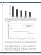
Figure 5. S-NACH treatment decreases sickling of red blood cells ex vivo under hypoxia. Total blood harvested from sickle cell disease mice (n=8) was mixed with a sulfated non-anticoagulant heparin derivative (S-NACH) at the dose of 1, 5, or 10 mg/mL and incubated in 2% O2 at 37°C for 1 h. Blood smears were made, stained, and the morphology of red blood cells (RBC) was analyzed. Hypoxia increased the percentage of sickled RBC. S-NACH treatment decreased the sickling of RBC in a dose-dependent manner. Phosphate-buffered saline (PBS) was used as a negative control and 5-hydroxymethyl-2-furfural (5-MF) was used as a positive control.
*P<0.05. SD: standard deviation
Figure 6. S-NACH treatment increases the survival of sickle cell disease mice under hypoxia. Sickle cell disease (SCD) mice were treated with phosphate-buffered saline (PBS, n=6) or 10 mg/kg sulfated non-anticoagulant heparin derivative (S-NACH, n=8). After 30 min, mice were incubated in a hypoxia chamber (5% O2), and the survival of animals was observed for 1.5 h. Surviving mice were euthanized, as per the guidelines. S-NACH treatment was associated with increased survival of mice.
S-NACH increased the levels of TFPI in plasma, decreased RBC sickling under normoxia and hypoxia, and reduced the levels of the pro-inflammatory mediators IL-1, IL-6, IFN-g, MCP-1, TNF-a, M-CSF, and VEGF while increasing anti-inflammatory factors such as IL-10, further establishing it as a promising bona fide multimodal candi- date drug worthy of additional investigations for acute and chronic disease management in SCD patients. In sum-
mary, our data demonstrate direct and support pleiotropic effects of S-NACH in ameliorating the complex patho- physiological mechanisms involved in SCD. Development into an effective drug would lead to improved outcome in patients globally with SCD, considering the current limit- ed therapeutic options, especially for the vast majority of patients with SCD who reside in underdeveloped areas of the world.52
538
haematologica | 2022; 107(2)
