
Page 77 - 2022_01-Haematologica-web
P. 77
Targeting AML MCL-1 re-sensitizes BCL-2 inhibition
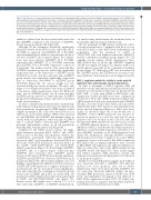
Figure 4. Mechanisms of synergy. Interactions of anti-apoptotic and pro-apoptotic BCL-2 proteins in control and BH3 mimetic-treated cells (A to D). OCI-AML3 cells were treated with venetoclax, AZD5991, or both (dose 1: venetoclax, 100 nM and AZD5991, 10 nM; dose 2: venetoclax, 250 nM and AZD5991, 25 nM) for 24 hours (h). (A to C) Interactions of BCL-2 (A), MCL-1 (B), or BCL-XL (C) with BIM, BAX, or BAK were determined by co-immunoprecipitation (co-IP) and western blot analysis. (D) Summary of the interactions. Roles of metabolic function and leukemia-stroma interactions (E and F). (E) OCI-AML3 cells were treated with venetoclax alone or in combination with IACS-10759, BL-8040, or AZD5991 with or without mesenchymal stromal cell (MSC) co-cultures for 48 h. Cell death in CD45+ cells were deter- mined by flow cytometry. (F) Migration (6 h) and adhesion (24 h) of BL-8040 treated-OCI-AML3 cells to MSC were measured. Migration to CXCL12 was used as a pos- itive control and random migration as a negative control. Experiments were performed in triplicates. Results are expressed as the mean ± standard error of the mean. IgG: immunoglobulin G.
similar for spleens from the mice treated with venetoclax plus AZD5991 compared to the untreated or AZD5991- treated groups (treatment d 25, Figure 6D).
Although all the treatments statistically significantly extended survival in the PDX model, venetoclax (54 d, P=0.0035), as expected, and AZD5991 (53 d, P=0.0081) showed minimal effects similar to that presented for the in vitro data. AZD4573, and to greater degrees the combina- tions, were more effective (AZD4573: 60 d, P=0.0002; venetoclax plus AZD4573: 71 d, P=0.0005; venetoclax plus AZD5991: 76.5 d, P=0.0005 compared to controls 51 d, Figure 6E). The median survival of the venetoclax plus AZD4573 treatment group was statistically significantly longer than that of the venetoclax or AZD4573 group (P=0.0004 for both), and the venetoclax plus AZD5991 group survival was statistically significantly longer than that of venetoclax (P=0.0004) or AZD5991 group (P=0.0012) (Figure 6E). No obvious weight loss was observed during the various treatments. The mice only began to lose weight several days before they succumbed to the disease (Online Supplementary Figure S5). Of the 60 mice, one in AZD5991 group, one in venetoclax plus AZD5991 group, and two in venetoclax plus AZD4573 group died of treatment procedures and were not included in the survival analyses.
In order to elucidate the treatment effects on phenotyp- ically-defined cell populations and on protein expression in these populations, we performed CyTOF analysis on BM samples (treatment d 25). Cells were clustered based on expressions of cell surface markers. While venetoclax had no, and AZD5991 and AZD4573 had minimal, effects in several AML cell populations, venetoclax plus AZD4573 and, to a greater degree, venetoclax plus AZD5991 had profound anti-leukemia activity in all cell populations including AML stem/progenitor cells (Figure 6F).
Compared to the vehicle treated controls, the single- agent treatments increased BCL-2, MCL-1, and c-MYC levels in most of the AML cell populations, whereas the combinations, particularly venetoclax plus AZD5991, decreased c-MYC in all populations, decreased BCL-2 in all but the CD34+CD38- population, decreased MCL-1 in the CD45+, CD34+CD38+CD123+, and CD34+CD38- CD123+ populations (Figure 7A). Interestingly, AZD5991, AZD4573, and, to a greater degree, venetoclax plus AZD5991 and venetoclax plus AZD4573 greatly decreased cell surface CXCR4 levels in all cell populations. AZD5991 and AZD4573 also decreased CD44 levels in all populations (Figure 7B). These results paralleled our in vitro findings suggesting that the inhibition of MCL-1 decreases CXCR4 and CD44 expression to suppress leukemia-stro- ma interactions. We also measured several signaling pro- teins and found that the combination treatments greatly decreased b-CATENIN and p-AKT in several AML cell populations (Online Supplementary Figure S6A), particularly in CD45+ and CD34+CD38-CD123+ cells (Figure 7C). Because mouse BM samples were collected 1, 3, or 2 d after venetoclax, AZD5991, or AZD4573 administration,
our analyses may underestimate the treatment effects on proteins/phosphor-proteins in leukemia cells.
We also performed CyTOF analysis on BM cells collect- ed from moribund mice. Compared with those in con- trols, BCL-2, MCL-1, and c-MYC levels in all leukemia cell populations, with the exception of c-MYC in CD34+CD38+ cells, were higher in venetoclax, AZD5991, and the combination treatment groups (Figure 7D). Cell signaling protein analysis (Online Supplementary Figure S6B) showed that in all but the AZD4573 treatment CXCR4 was suppressed (Figure 7E), whereas p-AKT was greatly induced especially in the combination treatment groups (Figure 7F). Although p-AKT was not induced in the AZD4573 group, the CXCR4 level recovered com- pared with that observed in the controls (Figure 7E and F).
MCL-1 regulates metabolic activity in acute myeloid leukemia cells and leukemia-stromal interactions
In order to determine if MCL-1 regulates AML cell metabolic activity and leukemia-stromal interactions inde- pendent of its anti-apoptotic functions, we knocked down BAX, BAK, or both with siRNA in OCI-AML3 cells. Suppression of BAX and BAK levels were confirmed by western blot 48 h after siRNA transfection (Figure 8A). siRNA transfected cells were then treated with AZD5991 (50 nM), a dose that did not affect viable cell count (Online Supplementary Figure S1E). Apoptosis, mitochondrial respi- ration, and cell migration/adhesion to MSC were deter- mined. As shown in Figure 8B, at the 50 nM dose of AZD5991, control siRNA-transfected cells showed no increase in apoptosis at 1 and 4 h, and only 5% more apoptotic cells over baseline at 24 h. The Bax, Bak, and Bax plus Bak siRNA-transfected cells were more resistant to AZD5991-induced apoptosis, as expected. However, inhibition of MCL-1 demonstrated the identical pattern of inhibition of mitochondrial respiration, detectable at 1 h after AZD5991 treatment, in all siRNA-transfected cells (Figure 8B). Furthermore, although AZD5991 treatment (24 h) induced low levels of apoptosis in control siRNA- transfected, but not in Bax and/or Bak siRNA-transfected OCI-AML3 cells, no apparent differences were observed in OCI-AML3 cell migration and adhesion inhibition to MSC in control, Bax, and/or Bak siRNA-transfected and AZD5991 treated cells (Figure 8C). These results suggest that MCL-1 regulates AML metabolic activity and leukemia-stromal interactions independent of its functions as an apoptosis regulator.
Finally, we treated AML patient samples (n=4) with low dose venetoclax (2.5 nM) or AZD5991 (12.5 nM) that induced low level apoptosis after 24 h (Online Supplementary Figure S7A) and found that AZD5991, but not venetoclax, effectively inhibited cellular and mito- chondrial ROS production, reduced CXCR4 and CD44 levels, and diminished cell migration and adhesion to MSC (Online Supplementary Figure S7B to D), further sup- porting that MCL-1 regulates mitochondrial respiration and leukemia-stromal-interactions in AML.
haematologica | 2022; 107(1)
69
