
Page 188 - 2022_01-Haematologica-web
P. 188
A. Hecht et al.
Mutational spectrum of CBL-mutated juvenile myelomonocytic leukemia and occurrence of secondary mutations
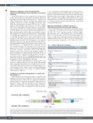
Clonal CBL mutations were confirmed for every patient in the cohort and were exclusively located in the linker and ring finger domains, as previously reported7,16,20 (Figure 1 and Online Supplementary Table S3). Twenty-eight patients had germline alterations of CBL, including six patients with deletions affecting these domains. All but two of these patients with germline CBL mutations showed a higher VAF of the same mutation in their tumor as a result of het- erozygosity (all VAF are shown in Online Supplementary Table S3). Of the two patients without heterozygosity, one had a deletion and the other a splice site mutation, compat- ible with previous reports in other patients.21 Interestingly, we identified five patients (15%) in our cohort with somat- ic-only CBL mutations. The somatic-only mutations were located at the same hotspot positions as those in the germline population and were homozygous in four patients and heterozygous in one patient. We then conducted fur- ther experiments in these five patients with somatic-only CBL mutations. To confirm that these mutations were in fact somatic in nature, buccal swabs were tested in four patients and a cord blood sample in the remaining patient. No exonic or intronic alterations of CBL or any other Ras pathway genes were found in the germline samples of these five patients by targeted deep sequencing. In one of the patients with a somatic-only CBL mutation, we discov- ered a heterozygous RUNX1 p.R166Q germline mutation, which has been reported in familial platelet disorder with propensity to myeloid malignancy syndrome.
Overall, missense CBL mutations at residue Y371 were the most common alteration in both groups. One patient with a germline CBL mutation was found to have a second- ary somatic CBL mutation with a lower VAF, leading to loss of heterozygosity. No other somatic secondary mutations were found in either cohort using our targeted 26-gene JMML panel at diagnosis.
Comparison of patients with germline or somatic-only CBL mutations
Table 2 describes the clinical presentation of patients with germline CBL mutations compared to those with somatic- only CBL mutations. There were no significant differences between the two cohorts in the patients’ characteristics at diagnosis (Table 2). Of note, patients in both cohorts pre- sented at a median age of 1 year. Clinical signs of dysmor- phia could not be used to distinguish between the groups,
as 13 of 20 patients with germline CBL mutations had no overtly syndromic features (65%, data not available for 8 patients). None of the patients with somatic CBL mutations presented with obvious signs of dysmorphia. In eight of 15 (53%) patients with germline CBL mutations for whom information was available, the mutation was documented to be inherited from a parent with equal rates of maternal and paternal inheritance.
Outcome and diversity of clinical courses of CBL-mutated juvenile myelomonocytic leukemia
The overall survival rate of the whole cohort after a medi- an follow-up of 3.7 years was 41% (95% confidence inter- val [95% CI]: 7-75%; median survival: 7.9 years) (Figure 2A). However, 45% of the patients (15 of 33) underwent HSCT at a median time of 0.5 years after initial diagnosis (range, 0.3-2.6 years). Overall survival of patients who underwent HSCT was 69% (95% CI: 36-87%; median sur- vival not reached) compared to 31% (95% CI: 1-74%;
Table 1. Patients’ characteristics at diagnosis.
Whole cohort (n=33)
Median age at diagnosis, years 1.1
Range
0.1-25.3
Gender
Male 14 Female 19
Median WBC at diagnosis, x109/L 34.2
Range
Median absolute monocyte count at diagnosis, x109/L 5.8
Range
Median platelet count at diagnosis, x109/L Range
Median hemoglobin at diagnosis, g/dL Range
Elevated hemoglobin F for age Abnormal cytogenetics Monosomy 7
Splenomegaly
Dysmorphic features present
Germline CBL mutation inheritance Maternal origin
Paternal origin
WBC: white blood cell count.
0.8-31.1
68 10-204
9.9 6.2-11.8
24% (6 of 25 with data available) 3% (1 of 30 with data available) 0
97% (29 of 30 with data available) 35% (7 of 20 with data available) 53% (8 of 15 with data available)
4 4
6.9-196.0
Figure 1. Germline and somatic CBL mutations in patients with juvenile myelomonocytic leukemia. Each dot represents one mutation found at the specified codon. The resulting change in amino acid is coded by colors. The stars represent six patients found to have deletions. 4H: four helix bundle; EF: EF-hand like domain; SH2: Src homology 2 domain; L: linker domain; RF: ring finger domain; Pro-rich: proline-rich domain; UBA/LZ: ubiquitin-associated/leucine zipper domain.
180
haematologica | 2022; 107(1)
