
Page 114 - 2022_01-Haematologica-web
P. 114
P. Kerbs et al.
A
B
D
C
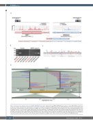
Figure 4. Detection and validation of the novel NRIP1-MIR99AHG fusion gene. Evidence for the NRIP1-MIR99AHG fusion gene in sample AM-0028-DX determined by various methods. (A) Schematic representation of the fusion transcript as predicted by RNA-sequencing. (B) Gel-electrophoresis of reverse transcriptase poly- merase chain reaction analysis of fusion breakpoint and NRIP1 exon 4. Three samples from cytogenetically normal patients with acute myeloid leukemia were used as negative controls. (C) A trace from Sanger sequencing of the fusion breakpoint. (D) Mapping of long reads from Nanopore sequencing of genomic DNA. Each line represents one read, which can be divided at the breakpoints of the fusion. Single parts of the read can be mapped to the positive strand (blue) at one locus with the other part mapped to the negative strand (red) at the other locus. The consensus inversed region is indicated by orange. The mapping structure of a highlighted read at the bottom shows that one part of the read was inversely mapped to the NRIP1 locus, while the other part was mapped to the MIR99AHG locus.
106
haematologica | 2022; 107(1)
