
Page 9 - 2021_12-Haematologica-web
P. 9
Images from the Haematologica Atlas of Hematologic Cytology: hemophagocytic syndrome
Rosangela Invernizzi
University of Pavia, Pavia, Italy
E-mail: ROSANGELA INVERNIZZI - rosangela.invernizzi@unipv.it
doi:10.3324/haematol.2021.279797
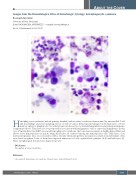
Hemophagocytic syndromes include primary (familial) and secondary conditions characterized by uncontrolled T-cell and macrophage activation and phagocytosis of cells of various hematopoietic lineages by the histiocytes of bone marrow, spleen or lymph nodes. Secondary forms can be reactive to an underlying infection, particularly in immuno- compromised hosts. They have also been reported in association with malignancies such as cancer and lymphomas. In this case of Epstein-Barr virus (EBV)-associated hemophagocytic syndrome, the bone marrow aspirate is slightly hypocellular and shows active hematopoiesis (A and B). Many histiocytes are scattered among the hematopoietic cells. They have a low nuclear:cytoplasmic ratio, an oval nucleus with a reticular chromatin pattern, inconspicuous nucleoli and abundant, often vacuolated cytoplasm. Some of them have ingested numerous red cells, erythroblasts, platelets (A-E) and leukocytes (F). These morphological features have diagnostic power.1
Disclosures
No conflicts of interest to disclose.
Reference
1. Invernizzi R. Hemophagocytic syndrome. Haematologica. 2020;105(Suppl 1):40-43.
ABOUT THE COVER
haematologica | 2021; 106(12)
3029
