
Page 55 - 2021_12-Haematologica-web
P. 55
IL1-IL1RAP axis in AML
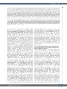
Figure 5. The IL1-IL1RAP signaling pathway affects normal hematopoiesis but not acute myeloid leukemia cell growth in the context of human mesenchymal stro- mal cells. (A) Growth curve of acute myeloid leukemia (AML) patient 1 (AML#1) CD34+ cells in liquid culture (left) and on a stromal layer of mesenchymal stromal cells (MSC) (right) ±IL1b in different concentrations. The arrow indicates from whereon IL1b was added. (B) Growth curve of CB CD34+ cells in liquid culture (left) and on a stromal layer of MSC (right) ±IL1b in different concentrations. The arrow indicates from whereon IL1b was added. (C) Schematic overview of experimental setup of co-cultures and conditioned medium (CM) transferring. (D and E) Growth curve (left) and cumulative cell number on day 14 (right) of CB CD34+cells in liquid (D) and on a stromal layer of MSC (E) with the addition of CM from a MSC culture (CM MSC), AML#1 CD34+ liquid culture (CM AML), AML#1 CD34+ MSC co-culture (CM MSC + AML), and AML#1 CD34+ MSC co-culture with the addition of 10 ng/mL IL1b (CM MSC + AML + IL1b). CM was added at day 4 (arrow) and every following demi-population. (F) Growth curve (left) and cumulative cell number on day 21 (right) of CD34+ peripheral blood stem cells (PBSC) grown on MSC with the CM from AML#18 (co-)cultures. Treatment conditions were similar to the once described in the legend of Figure 5D and E, adding one condition including AML CD34+ co-cul- ture with the addition of 10 ng/mL IL1b and 500 ng/mL Anakinra (CM MSC + AML + IL1b + Anakinra). CM was added at day 0 and at every following demi-population. (G) Colony-forming cell (CFC) assay of cord blood (CB) CD34+ treated with CM of IL1RAPhigh AML (AML#9 and AML#19) and IL1RAPlow AML (AML#20 and AML#21), which were cultured for 7 days on a MSC stromal-layer in the presence or absence of IL1b and Anakinra, before CM was harvested. Data of two biological duplicates are shown relative to the untreated condition. (H) Interleukin-1 receptor accessory protein (IL1RAP) expression on MSC measured by flow cytometry. (I) Quantitative real-time polymerase chain reaction of MSC stimulated with and without IL1b. Statistical analysis was performed using a Student’s t-test. (J) Cumulative cell number on day 21 of CD34+ PBSC grown on MSC (experimental setup identical to panel F) including conditions AML + IL1b and MSC + IL1b. Gartner’s and CM MSC + AML + IL1b (identical to panel F) has been added for direct comparison. (K) Growth curve (left) and cumulative cell number on day 11 (right) of CB CD34+ cells in triple co-culture with MSC and AML#16 CD34+ cells ±IL1b (L) Percentage of CB cells in triple co-culture with MSC and AML#22 ±IL1b at day 4. (M) Schematic model how AML cells might impact on normal hematopoiesis in the bone marrow niche, in part via the IL1-IL1RAP axis. Statistical analysis in all panels was performed using a Student’s t-test.* P<0.05; **P<0.01; ***P<0.001.
patients #10-13 and stimulated them with IL1b, FLT3L, SCF, IL3, or a combination thereof. Activation of signal transduction pathways was determined by western blot- ting for phosphorylated p65 (p-p65), phosphorylated p100 (p-p100), p52/p100, RelB, phosphorylated STAT5 (pSTAT5), phosphorylated p38 (p-p38), and phosphorylat- ed AKT (pAKT), (Figure 4D to G; Online Supplementary Figure S4B to E). Figure 4D to E illustrates AML patient #10 that expressed high levels of IL1RAP, FLT3, CD117 and CD123 within the CD34+ blast compartment. Stimulation with IL1b resulted in downstream activation of the canon- ical NFkB-(p65) and p38 pathways, whereas we observed limited activation of non-canonical NFkB (p52/p100, RelB, and p-p100), IL3 activated the STAT5 signaling pathway, FLT3L and SCF both activated the PI3K-AKT signaling pathway. Co-stimulation of IL1b with SCF resulted in a slightly increased downstream activation of the PI3K-AKT signaling, although no additive effects were seen on pSTAT5 or p-p38 (Figure 4D to E). In AML#13, which expressed IL1RAP, CD123 and CD117 and low levels of CD135, we observed that IL1b led to activation of p-p38 and a moderate activation of only canonical NFkB, IL3 induced pSTAT5, but again the IL3 signaling was not fur- ther potentiated by co-stimulation with IL1b (Online Supplementary Figure 4B to C). The third example (AML#11) showed increased p-p65 levels upon IL1b stim- ulation whereas the non-canonical NFkB was active at baseline but not further enhanced upon IL1b stimulation as read out by p52/p100 levels (Figure 4F to G). AML#11 responded to FLT3L, SCF and IL3 stimulation, but co-stim- ulation with IL1b did not further enhance activation of any of these signaling pathways (Figure 4F and G). The fourth patient sample (AML#12) had limited IL1RAP expression and showed no activation of neither canonical nor non-canonical NFkB upon stimulation with IL1b, no additional activation of the p-p38 signal that was already highly activated at baseline, and no effects of co-stimula- tion with IL1b were seen on IL3-induced pSTAT5 (Online Supplemental Figure S4D to E). Further evaluation of the non-canonical NFκB pathway showed that some primary AML patients already have high baseline non-canonical NFκB activity compared to THP1 cells. We did not observe differences in baseline non-canonical NFkB activ- ity in K562 IL1RAP+ and K562 IL1RAP- cells (Online Supplementary Figure S4F and G). Neither did we observe synergistic effects of IL1b with FLT3L or IL3 in THP1 cells on downstream phosphorylation of ERK (pERK), cJUN (p-
cJUN) and AKT (pAKT) (Online Supplementary Figure S4H, I, K and L). Addition of the anti-IL1RAP monoclonal anti- body (α-IL1RAP MAb), that could partly rescue IL1- induced upregulation of IL8 and CXCL1, did not alter phosphorylation levels of AKT and cJUN upon stimula- tion with IL1b in combination with FLT3L or IL3 (Online Supplementary Figure S4J to L). In summary, both the canonical NFkB and p38 pathway can be activated by IL1b in primary AML patients whereby downstream canonical NFkB signaling might be correlated to IL1RAP expression levels. We did not observe synergism of IL1b with other signaling molecules including FLT3L, SCF and IL3 on downstream phosphorylation of p65, p38, STAT5, AKT, ERK and cJUN.
The IL1-IL1RAP signaling axis reduces proliferation of normal hematopoietic cells but does not affect acute myeloid leukemia cell growth
We showed that IL1RAP is often upregulated in AML cells and that the receptor is functional, inducing an inflammatory gene expression signature. However, we found no evidence for a cell-intrinsic role for the IL1- IL1RAP axis in controlling cell proliferation or survival under stress conditions. Therefore, we wondered whether the inflammatory secretome induced via the IL1-IL1RAP signaling route plays a role in inducing an inflammatory BM niche that favors AML cell proliferation over normal hematopoiesis. Therefore, IL1RAP-expressing AML CD34+ cells were grown in liquid culture conditions or on MSC, in the absence or presence of IL1b. The addition of IL1b had limited impact on the proliferative capacity of primary AML CD34+ cells, neither when grown in liquid culture conditions nor when grown in co-culture with MSC (Figure 5A). Similarly, IL1b did not affect the prolif- eration of CB CD34+ cells when grown in liquid culture conditions, however, a marked, dose-dependent, reduc- tion in proliferation was noted when cultured on MSC (Figure 5B). From the MSC/AML cultures, the conditioned medium (CM) was harvested and transferred to CB- derived or PB-derived CD34+ cell cultures to determine the effects on proliferation and differentiation (Figure 5C). CM was harvested every time cultures were demi-popu- lated by taking a third of the volume and transferring it to CB- or PB-derived CD34+ cell cultures in a 1:1 ratio (v/v) (see the Online Supplementary Methods for more details)
The addition of the CM of AML#1 grown in liquid cul- ture conditions did not affect CB CD34+ growth, neither
haematologica | 2021; 106(12)
3075
