
Page 230 - 2021_12-Haematologica-web
P. 230
Case Report
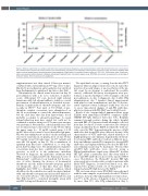
Figure 2. Dilution experiments on samples collected at day 1 post-admission (diagnosis), days 4 post-admission (after IVIg administration), day 7 post-admis- sion (marked clinical improvement with platelet count normalization) and day 15 post-admission (deterioration of patient’s status and death). Dilutions were made in normal heated plasma (containing normal immunoglobulin G [IgG] level) or in modified Tyrode’s buffer at 1/10, 1/20, 1/40 dilution ratios. Platelet acti- vation was assessed without heparin in platelet-activating anti-platelet factor 4-serotonin-release assay (PF4-SRA) and results are expressed in percentage of serotonin release. IVIG: intravenous immunoglobulin.
supplementation was then started (2 liters per minute) combined with oral moxifloxacin 400 mg od for 5 days. On day 12 post-admission, anticoagulation was switched from fondaparinux to apixaban 5 mg twice a day (bid).
Unfortunately, the clinical status worsened on day 12 post-admission with a de novo reduction of platelet count. Abdominal CT scan showed right adrenal hematoma with left adrenal infiltrate, which is a usual presentation of adrenal infarction, as described in auto- immune heparin-induced thrombocytopenia and also recently in VITT.4,5 Four units of 500 IU/mL of pro- thrombin complex concentrate were administered on day 14 as an attempt to control the adrenal hematoma, but she died later that day from hypovolemic shock probably secondary to adrenal hemorrhage. A causal adrenal infarction may have existed but could not be confirmed as neither an injected CT scan nor an autopsy was performed. Nevertheless, adrenal insufficiency was not documented and cortisol levels on day 14 was still in the upper range (i.e., 21 mg/dL, normal range: 6.2-18 mg/dL). Moreover, it must be noted that although apixa- ban was last administered on day 13 in the morning and was never reintroduced, its plasma level on day 14 was 353 ng/mL (usual Ctrough range: 22-177 ng/mL) and was still 132 ng/mL on day 15. The accumulation of apixaban may thus have contributed to, or even trig- gered, this bleeding event. The initial infusion of platelet concentrates may also have contributed to disease pro- gression, as well as to the pulmonary embolism observed at diagnosis. An immediate treatment with IVIg, as now recommended by the American Society of Hematology,6 could have been beneficial but the pres- ence of active bruising, the absence of documented thrombosis and the marked thrombocytopenia guided our therapeutic choice at that time. This case further highlights how VITT is a dynamical condition, which should not be discounted in a recently vaccinated patient with only thrombocytopenia and increased D-dimer lev- els, even in the absence of documented thrombosis, and that IVIg should be considered promptly, along with anti-PF4 testing and thrombosis screening.
The rapid fatal outcome, occurring 2 weeks after VITT diagnosis while an improvement was noticed, raised the question of an early relapse or an exacerbation of the ini- tial event. In an attempt to understand the possible cause(s), additional laboratory investigations were per- formed, as reported in Figure 1. Blood samples collected post-admission on day 1 (diagnosis), day 4 (after IVIg administration), day 7 (marked clinical improvement with platelet count normalization) and day 15 (deterio- ration of patient’s status, leading to death) were selected to assess time-related changes of anti-PF4 antibodies, platelet activation with PF4-SRA7 and total IgG levels.
Result of the enzyme-linked immunosorbent assay (ELISA) with immobilized PF4/PVS complexes (LIFE- CODES PF4 IgG, Immucor Lifecodes, Jette, Belgium) remained strongly positive during the whole hospital stay with OD >3.00 measured with all samples collected from day 1 to day 15. Positive results were also obtained using a modified in-house ELISA in which the wells are only coated by PF4,8 demonstrating the presence of IgG antibodies that bound PF4 alone (data not shown). Such characteristics of VITT antibodies, shared with those of highly pathogenic auto-immune heparin-induced throm- bocytopenia (HIT) antibodies,9 indicate a different speci- ficity and affinity towards PF4 compared to classical HIT antibodies, and explain why their detection by HIT-ded- icated immunoassays may be inadequate.7 The fact that anti-PF4 IgG antibodies were detected using a PF4/polyvinylsulfonate rapid assay (HemosIL® AcuStar HIT IgG assay) is particular since this assay failed to detect anti-PF4 antibodies in most of the reported VITT cases. Nevertheless, the HemosIL® Acustar HIT IgG assay rarely gives weak positive results in patients with likely VITT diagnosis, as stated by Platton et al., who reported two positive results out of 31 patients, all of whom had anti-PF4 IgG detected by ELISA.10
PF4 serotonin-release assay (PF4-SRA) was performed with the same samples, as previously described.7 Platelet activation, measured through maximal serotonin release, was 90% in the absence of heparin on day 1, decreased by more than 50% after IVIg administration (day 4,
3250
haematologica | 2021; 106(12)
