
Page 138 - 2021_12-Haematologica-web
P. 138
M. Nairz et al.
E
F
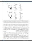
Figure 5. Cell-type specific effects of Hfe on protein expression in the spleen. Spleen homogenates were prepared to quantify the expression of iron and immune relevant proteins by enzyme-linked immunosorbent assay. Protein levels of Fpn1 (A), H-Ft (B), Tfr1 (C), Lcn2 (D), IFN-g (E) and Nos2 (F), normalized for total protein content, are depicted as means ± standard deviations. Statistically significant differences as calculated by unpaired, two-sided student t-test are indicated. n=10 for Hfe+/+, n=12 for Hfe-/-, n=20 for AlfpCre– Hfefl/fl, n=14 for AlfpCre+ Hfefl/fl, n=15 for LysMCre– Hfefl/fl, n=15 for LysMCre+ Hfefl/fl.
contributed to the insufficient induc mice is linked to increased cellular iron levels, we next maintained WT mice on an iron adequate (IA) or high iron diet for three weeks to induce iron overload (IO) prior to Salmonella infection. We observed an increased bacterial load in the serum (Figure 6D), spleen and liver (Online Supplementary Figure S5B and C) along with unaltered IL-6 (Figure 6E) but reduced IFN-g concentrations in the serum (Figure 6F). This finding suggests that reduced levels of IFN-g, the cen- tral cytokine orchestrator of immune responses against intracellular bacteria,28 are a direct consequence of increased serum iron.
Discussion
The challenge of mice with the facultative intracellular bacterium S. Tm. uncovered an important extra-hepatic function of Hfe in macrophages and novel cell type-specif- ic roles of Hfe in infection control and immune regulation: mice lacking Hfe either in all cell types or selectively in the myeloid compartment were more resistant to Salmonella infection and protected from early death compared to WT littermates expressing Hfe. Conversely, hepatocyte-specif- ic Hfe deletion was deleterious to the host, triggering early death in response to Salmonella infection. These findings are somewhat unexpected because the primary iron over- load patterns of Hfe-/- mice and hepatocyte-specific Hfe
knockout mice are alike.25 Mice with myeloid-specific Hfe deletion by contrast, show no apparent iron-phenotype but are resistant to S. Tm. infection much like Hfe-/- mice.13 This suggests that the putative immune-regulatory roles of Hfe in macrophages are partially separated from its iron-regulatory functions or mediated via micro-environ- mental rather than systemic effects. Further studies using combinations of pathogens and exogenous iron sources will be required to ravel out underlying regulatory net- works. However, the lack of Hfe in macrophages is suffi- cient to explain the improved survival of constitutive Hfe-/- mice infected with Salmonella. This fact may directly be related to the profound tropism of Salmonella for myeloid cells and points to an important Hfe function in macrophages.29,30 We noted that upon Salmonella infec- tion, LysMCre+ Hfefl/fl and Hfe-/- mice have lower iron levels in the spleen. Indeed, it has been suggested that lower lev- els of iron in macrophages may be protective against pathogens such as Salmonella that propagate within macrophages.31
Surprisingly, we found accelerated death of AlfpCre+ Hfefl/fl mice due to impaired resistance to Salmonella infection, although macrophage iron content was unaffected, and bacterial loads in the spleen and liver were not different as compared to AlfpCre– Hfefl/fl mice. Our data rather indicate that enhanced extracellular bacterial proliferation in the iron-rich serum is deleterious to AlfpCre+ Hfefl/fl mice. Apart from iron-induced bacterial growth, the reduced levels of
3158
haematologica | 2021; 106(12)
