
Page 99 - 2021_10-Haematologica-web
P. 99
CDK4 or CDK6 deletion in adult hematopoiesis
Figure S3E). These results underscore the dominance of CDK6 for the maintenance and proliferation of the stem cell pool.
CDK4 and CDK6 effect the myeloid and lymphoid pro- genitor pool
In complete Cdk6-/- mice, myeloid-primed progenitors (Lin-c-Kit+Sca-1-; LKS-) and their subpopulations including common myeloid progenitors (CMP), megakaryocyte/erythroid progenitors (MEP) and granulo- cyte/macrophage progenitors (GMP) are slightly increased.18 Three weeks upon deletion of CDK6 in adult hematopoiesis, the myeloid-primed progenitor pool remained unaltered in Cdk6D/D BM as compared to litter- mate controls (Figure 4A; Online Supplementary Figure S4A
AB
and B). Myeloid-primed progenitors accumulated in Cdk4D/D BM (Figure 4A) as did CMP (LKS- CD34+CD16/32lo), GMP (LKS-CD34+CD16/32hi) and MEP (LKS-CD34-CD16/32lo) (Online Supplementary Figure S4B). In contrast, 6 weeks after Cdk4 or Cdk6 deletion, the per- centage of myeloid progenitors returns to an unaltered state in Cdk4D/D compared to controls, but increased in Cdk6D/D (Online Supplementary Figure S4G) reflecting the Cdk6-/- phenotype. Independent of deletion span CLP (Lin- IL-7R+c-KitmidSca-1mid) are enriched only in Cdk6D/D BM (Figure 4B; Online Supplementary Figure S4A and G) accompanied by elevated numbers of differentiated CD3+ T cells (Figure 4D; Online Supplementary Figure S4C and H) and unchanged numbers of CD19+ B cells (Online Supplementary Figure S4C to D). Despite the lack of any
CD
E
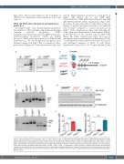
Figure 1. Reduced CDK4 or CDK6 expression in Cdk4fl/fl or Cdk6fl/fl Mx1-Cre mice. (A) The presence of loxP-flanked Cdk4 (left) or Cdk6 (right) were confirmed by poly- merase chain reaction (PCR) on genomic DNA of murine tissue, 100 bp marker. (B) Treatment regime for Mx1-Cre mediated deletion of Cdk4 (Cdk4∆/∆) or Cdk6 (Cdk6∆/∆) by polyinosinic–polycytidylic acid (poly(I:C)) injection (200 mg, intraperitoneally, three times every 3 days). (C) Deletion PCR: confirmation of Cdk4∆/∆ (top) or Cdk6∆/∆ (bottom) DNA in splenocytes showing wild-type (wt), floxed and delta bands. Analysis was performed 3 weeks post final poly(I:C) injection. (D) Immunoblotting: protein levels of CDK4 and CDK6 in spleen cells (Cdk4fl/fl or Cdk6fl/fl, Cdk4∆/∆, Cdk6∆/∆, n=3/genotype). α-tubulin served as a loading control. Analysis was performed 3 weeks post final poly(I:C) injection. A representative blot of at least three independent experiments is shown. (E) Quantification of immunoblot shown in (D), signal intensities were normalized to α-tubulin levels (A.U. [arbitrary units]). Cdk4∆/∆ or Cdk6∆/∆ protein levels were compared to the respective controls by performing Mann- Whitney U tests.
haematologica | 2021; 106(10)
2627
