
Page 9 - 2021_10-Haematologica-web
P. 9
Images from the Haematologica Atlas of Hematologic Cytology: trypanosomiasis
Antonello Malfitano1 and Rosangela Invernizzi2
1IRCCS Policlinico San Matteo Foundation and 2University of Pavia, Pavia, Italy E-mail: ROSANGELA INVERNIZZI - rosangela.invernizzi@unipv.it
doi:10.3324/haematol.2021.279412
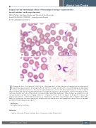
Following the bite of an infected TseTse fly or a diatomine bug, for the African or American species respectively, Trypanosoma spp. parasitizes the peripheral blood, where it is found outside red blood cells. Identifying morphological features of trypomastigotes (i.e., the parasite forms that may be observed in the peripheral blood) are the elongated shape, the flagellum and the undulating membrane. The flagellum originates in the kinetoplast, a DNA-containing structure, and runs alongside the body of the parasite, enveloped in the undulating membrane. These features are common to both African variants, responsible for sleeping sickness, i.e., Trypanosoma brucei rhodesiense (Figure A, B, thin blood smear) and Trypanosoma gambiense, and the American species, i.e., Trypanosoma cruzi which causes Chagas disease (Figure C, thick film). Only for Trypanosoma cruzi have amastigotes been described in bone marrow and lymph node smears; these are indistin- guishable from Leishmania amastigotes.1
Disclosures
No conflicts of interest to disclose.
Reference
1. Malfitano A, Invernizzi R. Parasitic and fungal diseases. Haematologica. 2020;105(Suppl 1):29-39.
ABOUT THE COVER
haematologica | 2021; 106(10)
2537
