
Page 261 - 2021_10-Haematologica-web
P. 261
Case Reports
ABC
DEF
GH
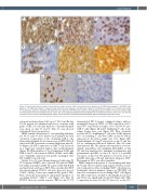
Figure 2. Immunohistochemical stains of post-chimeric antigen receptor (CAR) T-cell relapse tumor specimens. (A) CD19 stain, patient 1*; (B) CD20 stain, patient 1*; (C) CD3 stain, patient 1, showing T cells within the tumor specimen; (D) PDL1 stain, patient 1; (E) CD19 stain, patient 2*; (F) CD20 stain, patient 2*; (G) CD3 stain, patient 2, showing T cells within the tumor specimen; (H) PDL1 stain, patient 2. All images were taken at 1000x.*This work is licensed under a CC-BY Creative Commons attribution license, version 4.0. It was originally published in Nature Medicine and is attributed to Nirav N. Shah et al., and the orig- inal version can be found at the following URL: (https://www.nature.com/articles/s41591-020-1081-3). Minor formatting changes were made.
retroperitoneal mass from 9.4x9 cm to 7.5x9.4 cm. On day 52, the patient was admitted with fever, consistent with grade 1 CRS. A second and third dose of pembrolizumab were given on days 60 and 81. Day 93 scans showed widespread disease progression.
PBMC samples from patient 1 corresponding with days 28, 38, 60 and 90 were thawed and analyzed by flow cytometry. Composition of the circulating CAR T cells after pembrolizumab showed an increase in CD8:CD4 cell ratios and CAR T persistence remained high from days 28- 90 (Figure 1A). PD-1 expression on CAR T cells decreased in a rapid and sustained fashion after pembrolizumab (Figure 1B). Other markers of T-cell exhaustion (LAG3 and TIM-3) and T-cell activation (CD40L, IL2RA, and 4-1BB) were minimally expressed and greatly unchanged after PD-1 inhibitor (not shown).
Patient 2 is a 69-year-old-man diagnosed with stage IV TP53-mutated DLBCL who achieved CR with R-CHOP but within 2 months had PD. He achieved CR with R-ICE and underwent autologous HCT. Three months after HCT the patient relapsed. LV20.19 cells were given at a dose of 2.5x10⁶ cells/kg. Course was complicated by grade 1 CRS. Flow cytometry demonstrated exponential expansion of circulating LV20.19 CAR T cells (Figure 1C). Day 28 PET/CT showed a partial response (PR) but day 71 CT
demonstrated PD. A biopsy confirmed relapse with no meaningful change in CD19 or CD20 expression (100% and 100% respectively at relapse; 100% and 99.5% pre- CAR T cells) (Figure 2E and F). Infiltrating T cells in the relapse biopsy were seen (Figure 2G). Flow cytometry revealed 18.5% of infiltrating T cells were CAR+ whereas 26% of circulating T cells were CAR+ at day 80.2 PD-L1 expression in the tumor sample was 1-5% (Figure 2H). Pembrolizumab 200 mg IV was given on day 73 and day 100 in combination with neck radiation. Day 120 scans demonstrated PD. The patient subsequently received rit- uximab, gemcitabine and oxaliplatin (R-GemOx) on day 127 and was transitioned to rituximab, bendamustine and polatuzumab (pola-BR) on day 141. Cycle 2 of pola-BR was given on day 162. The patient received six cycles of pola-BR achieving a CR and underwent allogeneic HCT. He remains in CR 1 year after transplant.
PBMC samples from patient 2 corresponding with days 58, 78 and 118 were thawed and analyzed by flow cytom- etry. Composition of the circulating CAR T cells after pem- brolizumab showed minimal changes in CD8:CD4 cell ratios but a sustained rise in circulating CAR T cells (Figure 1C). PD-1 expression on CAR T cells initially decreased at day 78 but rose significantly by day 118 (Figure 1D). TIM- 3 expression decreased at day 78, then rose at day 118.
haematologica | 2021; 106(10)
2789
