
Page 242 - 2021_10-Haematologica-web
P. 242
Letters to the Editor
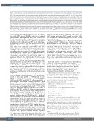
Figure 1. Cytomegalovirus reactivation impacts T-cell reconstitution following haploindentical hematopoietic stem cell transplantation with post-transplant cyclophosphamide. (A) Integrated median fluorescence intensity (iMFI) values, considering both expression level and frequency of marker+ cells, of the CD8+ or CD4+ T-cell PhenoGraph clusters were visualized by heatmaps, while PhenoGraph cluster dynamics (median percentages of total CD8+ or CD4+ T cells) in hap- loidentical hematopoietic stem cell transplantatation (haplo-HSCT) patients were revealed by balloon plots. (B) Principle component analysis (PCA) of CD8+ and CD4+ T-cell cluster frequencies. The left panel depicts the clusters driving the PCA, the central and right panel show the median PCA coordinates of all patients, grouped together or according to cytomegalovirus (CMV) viremia (subclinical viremia, n=6; clinical viremia, n=13; no viremia, n=2), per time point in months after transplant. The central and right panel should be read in conjunction with the left panel; the relative position of data points in the central and right panel is indicative of dominance of T-cell clusters shown with a similar relative position in the left panel. (C) Dynamics of CD8+ TEMRA, CD4+ TTE TH1 and CD8+ naïve clusters in haplo-HSCT patients experiencing post-transplant CMV viremia. Medians with the number of patients per time point are shown and error bars repre- sent interquartile range. Significance was determined by Kruskal-Wallis test and P-values are shown at the upper border of the plot for each time point (*P<0.05). (D) Flow cytometric analysis of CD45RA and T-bet expression in CD8+ and CD4+ T cells at month 3 and month 6-8 after transplantation. For both CMV groups a single representative patient is shown. (E) Hierarchical metaclustering using the Ward minimum variance method, of haplo-HSCT patients per month, based on the frequency of CD8+ and CD4+ T-cell PhenoGraph clusters. aGvHD: acute graft-versus-host disease; Exh: exhausted; PB: peripheral blood; Prolif: proliferating; TCM: central memory T cell; TEF: effector T cell; TEM: effector memory T cell; TEMRA: effector memory re-expressing CD45RA T cell; TM: memory T cell; TREG: regulatory T-cell T cell; TTE: terminal effector T cell; TM: transitional memory T cell.
effector phenotypes expressing T-bet, often in conjunc- tion with CD57 and/or CD45RA, indicative of terminal differentiation. Although CD8+ and CD4+ CMV-specific T-cell immunophenotypes displayed similar dynamics, we also observed lineage-specific differences, including identification of multifunctional CD4+ cluster 4 that high- ly expressed IL-2, IFN-γ, TNF and intermediate levels of CD107a. CMV-specific CD8+ T cells rarely expressed IL-2, rather, they commonly expressed IFN-γ and TNF. IFN-γ–TNF+ CD8+ T cells formed a minority, whereas IFN- γ+TNF+ T cells were commonly seen and dominated the overall response from month 5 onwards. Interestingly, we identified CD4+ clusters 3 and 11 expressing high lev- els of CD107a, T-bet, IFN-γ, TNF and CD57, reminiscent of killer-like cells otherwise identified in the CD8+ com- partment. Overall, the CMV-specific T-cell response showed maturation of effector functions over time (acquisition of at least three functions simultaneously), most prominently among CD8+ T cells (Figure 2C). The frequency of both CD8+ and CD4+ CMV-specific T cells in the PB of haplo-HSCT patients was greatly increased compared to that of the graft and PB of CMV-seropositive donors or PB of unrelated healthy controls, but pheno- type distribution at 1 year was remarkably similar, sug- gesting re-establishment of physiological homeostasis (Figure 2D).
We next asked whether control of CMV viremia is associated with a greater abundance, or a specific func- tional or phenotypic profile, of CMV-specific T cells. Huntley et al. reported >1 and >1.2 counts/mL of IFN-γ+ CMV-specific CD8+ and CD4+ T cells, respectively, to protect against reactivation following haplo-HSCT with pt-cy,13 but other studies, predominantly on HLA- matched HSCT recipients, reported that multifunctional responses have a stronger predictive value.14,15 We did not detect a significant difference in the total count of CMV-specific CD8+ or CD4+ T cells in the first 6 months post-transplant between patients with subclinical versus clinical CMV viremia, although CMV-specific CD4+ T cells tended to be present in higher amounts among patients with subclinical CMV (Figure 3A). Significant differences may occur at later time points, but our analy- sis was limited by the low number of patient samples at these time points. Analyzing the dynamics of each T-cell cluster separately, we found lower counts of multifunc- tional (cluster 4), proliferating (sum of phenotypically similar clusters 8 and 10) and TTE TH1 (sum of phenotyp- ically similar clusters 2 and 9) CD4+ T cells at month 3-4 for patients with clinical viremia (Figure 3B). Although the size of each given patient group is low, patients with repeated CMV episodes requiring multiple treatment cycles tended to develop even lower counts of these T- cell phenotypes (Online Supplementary Figure S2B). No such trends were seen in the CD8+ T-cell compartment
(data not shown), thereby suggesting that control of CMV viremia post-haplo-HSCT mainly associates with the development of distinct antigen-specific CD4+ T-cell immunophenotypes.
In conclusion, CMV-specific T cells were primed early after haplo-HSCT with pt-cy and initially displayed a proliferating/activated phenotype, that was quickly replaced by a terminal effector phenotype. One year after transplant, CMV-specific T-cell profiles were similar to those of the CMV-seropositive donor, suggesting re- establishment of physiological homeostasis. Uncontrolled viral replication associated with lower abundance of distinct CMV-specific CD4+ T-cell immunophenotypes, hinting at a possible role of these cells in CMV control following haplo-HSCT with pt-cy. These data require additional, future investigations for confirmation.
Jasper J. P. van Beek,1* Alessandra Roberto,1* Simone Puccio,1 Federica De Paoli,1 Giulia Graziano,2 Elisa Salviato,2
Giorgia Alvisi,1 Veronica Zanon,1Alice Scarpa,1 Elisa Zaghi,1 Michela Calvi,1 Clara Di Vito,3 Rossana Mineri,1 Barbara Sarina,1 Chiara De Philippis,1 Armando Santoro,1,4 Jacopo Mariotti,1 Stefania Bramanti,1 Francesco Ferrari,2,5 Luca Castagna,1 Domenico Mavilio1,3 and Enrico Lugli1
1IRCCS Humanitas Research Hospital, Rozzano, Milan; 2IFOM, the FIRC Institute of Molecular Oncology, Milan; 3Department of Medical Biotechnologies and Translational Medicine (BioMeTra), University of Milan, Milan; 4Department of Biomedical Sciences, Humanitas University, Pieve Emanuele, Milan and 5IGM-CNR, Institute of Molecular Genetics "Luigi Luca Cavalli Sforza",
National Research Council, Pavia, Italy
*JJPvB and AJ contributed equally as co-first authos Correspondence:
ENRICO LUGLI - enrico.lugli@humanitasresearch.it doi:10.3324/haematol.2020.276352
Received: November 17, 2020.
Accepted: June 16, 2021.
Pre-published: July 8, 2021.
Disclosures: EL receives reagents in kind from BD Biosciences Italy as part of a collaborative research agreement, and preclinical funding from Bristol-Myers Squibb on topics unrelated to the content of this manuscript; AS has received honoraria as advisory board member from Bristol-Myers Squibb, Servier, Gilead, Pfizer, Eisai, Bayer and Merck Sharp & Dhome; as speaker’s bureau member from Takeda, Roche, Abb-vie, Amgen, Celgene, Astrazeneca, Lilly, Sandoz, Novartis, Bristol-Myers Squibb, Servier, Gilead, Pfizer, Arqule, Eisai; and for consultancy from Arqule. The other authors declare no financial interests.
Contributions: JJPvB, AR and EL conceived the study; FDP, GA, VZ, AS, EZ, MC and CDV performed the experiments; RM, BS,
2770
haematologica | 2021; 106(10)
