
Page 222 - 2021_10-Haematologica-web
P. 222
Letters to the Editor
IRAKi to the cell cultures (Figure 2C). In line with the increased blockade of NF-κB signaling, the addition of CPI203 synergistically improved IRAKi cytostatic effect in the three cell lines, as attested by an 86% blockade in cell proliferation, significantly higher than the 19% activity achieved by IRAKi alone (combination index [CI]: 0.52, Figure 2D). Importantly, the co-operation between the IRAKi and CPI203 involved a remarkable downregulation of MCL-1 (Figure 2C), which was accompanied by a 36% increase in the relative apoptosis rate when compared with IRAKi and CPI203 used separately (Figure 2F).
In order to further validate the activity of the drug com- bination, primary lymph node biopsies from DLBCL patients with either MYD88wt or MYD88 L265P were co- cultured in the presence of a feeding stromal monolayer as previously reported,12 and treated with the different drugs as above. While IRAKi-CPI203 was almost inactive in MYD88wt cells, the combination induced a 16% augmenta- tion in relative apoptotic cell death in the MYD88 L265P primary co-culture (Figure 3A, left panel), which was accompanied by a 12% decrease in the fraction
of cells with high contents of IL6 mRNA, a percentage superior to what observed upon treatment with each drug alone (Figure 3B, right panel).
Among the above mentioned genes, CD44 expression and IL-6 serum levels have been described as prognostic markers in DLBCL.13,14 In order to investigate the role of these two factors in the response of ABC-DLBCL cell lines to IRAKi-based treatment, HBL-1 and OCI-LY3 cells were stimulated with 0.5 mM of the CD44 ligand, hyaluronic acid (HA), or exposed to a 5 mg/mL dose of the IL-6 block- ing antibody tocilizumab, prior to a 72-hour treatment with the drugs. In the case of HA, cells were exposed to IRAKi (50 mM) +/- CPI203 (0.5 mM), while effect of tocilizumab pretreatment was evaluated in IRAKi-treated cells. Cell response was determined by fluorescence microscopy recounting of cells with high contents in F- actin and by MTT assay, respectively. As shown on Figure 3B, both IRAKi and CPI203 were able to block actin poly- merizationby50.4%and54.5%,respectively,whilethe drug combination achieved a total 77.9% decrease in cells with high contents in F-actin following stimulation of
AB
C
D
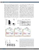
Figure 1. Limited activity of IRAKi single agent in activated B-cell - diffuse large B cell lymhoma cell lines with MYD88 L265P in relation with incom- plete inhibition of NF-κB gene signatures. (A) MTT assay showing that IRAKi (50 mM) elicited a partial and transitory response in activated B-cell - diffuse large B cell lymhoma (ABC-DLBCL) cell lines, while germinal center B-cell (GCB)-DLBCL cell lines were almost completely resistant to the compound. (B) IRAKi efficiently blocked the phosphorylation of IRAK1 at Thr29 and IRAK4 at Thr345, in the three ABC-DLBCL cell lines with MYD88 L265P. b-actin was used as a loading control. (C) Gene expression signatures of NF-κB in HBL-1, OCI-Ly3 and OCI-Ly10 cell lines exposed to IRAKi as above, highlighting that IRAKi treatment slightly affects this pathway (note that only 2 out 3 gene sets showed a false discovery rate [FDR] below 0.05). NES: normalized enrichment score. (D) Quantitative reverse transcriptase polymerare chain reaction (RQ- PCR) analysis of the predominant NF-κB-regulated genes IL6, IL10, CCL3, and IRF4, and the IRAK1/4 target gene MCL-1 in ABC-DLBCL cell treated by IRAKi as before (*P=0.01; **P<0.001).
2750
haematologica | 2021; 106(10)
