
Page 219 - 2021_10-Haematologica-web
P. 219
Letters to the Editor
characterized by a low socio-economic level (unavailable public data to date). Notwithstanding, this rather high rate of seropositivity shows that SCD children were actu- ally exposed to SARS-CoV-2 and presented indeed a benign clinical course, comparable to the general popula- tion of the same age.
This finding was somewhat puzzling given the delete- rious inflammatory potential of SARS-CoV-2 infection and the involvement of inflammation in the pathobiolo- gy of SCD. An uncontrolled multifactorial inflammatory response (notably via activation of neutrophils and platelets) is responsible for both acute vaso-occlusive events (VOE) and longer-term organ damage. Importantly, children with SCD are, like their adult coun- terparts, susceptible to viral factors triggering the occur- rence of ACS.4
One explanation may pertain to a modified IFN basal status in SCD. Recently, it was shown that homozygous SCD patients had an increased basal high plasmatic IFN- α concentration associated with an abnormal activation of the IFN-I signaling pathway in neutrophils.10 Given that during SARS-CoV-2 infection, an increased IFN-I plasmatic level contributes to control viral load by regu- lating the expression of the cellular entry receptor for SARS-Cov-2 (ACE2), we sought to further explore the
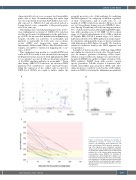
A
potential protective role of this pathway, by analyzing the IFN-I signature11 in a subgroup of children, regardless of their seroprevalence and at steady state (i.e., >2 months of a VOE or infectious episode). Of note, we did not test this pathway during an acute COVID-19 infec- tion. Among 25 randomly selected homozygous patients, 80% of them (n=20) had an abnormally high IFN-I signa- ture, with a median score of 11.3 (IQR, 3.2-32.9, normal range, <2.3) and a high plasmatic level of IFN-α (median 40.6 fg/mL; IQR, 22.4-64.4, normal range, <0.1), indicat- ing basal activation of the IFN-I pathway in a large major- ity of patients, independently of age or chronic treatment with hydroxyurea (HU) (Figure 1A and B). We found no statistical correlation between the IFN-I signature and seroprevalence.
A number of factors specific to SCD may trigger IFN-I and explain its activation at steady state. Chronic hemol- ysis, for instance, generates circulating cell-free heme, which is a major erythrocyte damage-associated molecu- lar pattern (DAMP) susceptible to trigger activation of the IFN-1 pathway. DAMP along with reactive oxygen species generation, Toll-like receptor 4 activation, neu- trophil extracellular trap generation, DNA, and other unknown factors contribute to the baseline sterile inflam- mation in SCD by activating the inflammasome pathway
C
B
Figure 1. Interferon-I signature and kinetics of SARS-CoV-2 serology in sickle cell disease patients. (A) Interferon-I (IFN-I) signature in 25 sickle cell disease (SCD) patients according to age. IFN-I signature was considered increased above a score of 2.3. (B) Basal IFNα concentration in plasma from healthy donors (AA, n=34) and SS patients (SS, n=25) using a digital enzyme-linked immunsorbent assay (SIMOA). (C) Evolution of serological titer (immunoglobulin G [IgG]) in 17 SCD patients following the first wave of the SARS- CoV-2 epidemic. The different zones represent the level of IgG titers and interpretation: grey: negative IgG titers; yellow: intermediate IgG titers; red: pos- itive IgG titers.
haematologica | 2021; 106(10)
2747
