
Page 109 - 2021_10-Haematologica-web
P. 109
Secreted MEF factors maintain HSC cultures
generation of the donor pool of short- and LTR cell- enriched fractions of CD34+ and CD34- CD48- LSK cells (Figure 3G, Online Supplementary Figure S4B, C), respective- ly. However, the total number of CD34- CD48- LSK cells was higher in mice receiving cells from MEF-CM 2GF cul- tures than in those receiving cells from MEF-CM 4GF and SFM 4GF cultures (Figure 3G).
Discussion
Some of the biggest challenges in hematology are to understand how niche cells regulate self-renewal and sur- vival of HSC and to develop methods with which HSC can be robustly and reproducibly expanded in vitro. In the cur- rent work, we used MEF as a source of secreted HSC-regu- latory molecules and found that CM prepared from these stromal cells promoted the survival and self-renewal of
AB
CD
murine HSC in culture. MEF are derived from primary material, which can be easily generated, even from most mouse mutants causing embryonic lethality, and used by many investigators to study mechanisms of embryonic stem cell regulation. Here, we demonstrate that MEF are a robust alternative in protocols aimed at maintaining and expanding HSC. Furthermore, MEF can be used to identify secreted factors involved in HSC maintenance. We show that MEF-CM not only supports the survival of HSC- enriched CD34- SLAM cells in cultures supplemented with 2GF, but also improves their self-renewal under both 2GF and 4GF conditions.
The additional use of MEF-CM prepared under serum- free conditions improves survival and self-renewal of CD34- SLAM cells. In this respect, MEF-CM performs the same survival-promoting function as the CM from the stro- mal cell line UG26-1B6 that we studied previously.1 As in our previous study, the stromal CM did not seem to affect
EFG
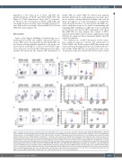
Figure 2. Maintenance of LSK cells and differentiation of CD34- SLAM cells in single cell cultures. Cells were cultured in either SFM 4GF, MEF-CM 4GF, or MEF-CM 2GF as indicated. (A) Representative flow cytometric plots of annexin V and propidium iodide (PI) staining of sorted LSK cells cultured for 48 h. (B) Percentage of viable (annexin V- PI-) and apoptotic (annexin V+) LSK cells after 48 h of culture. (C) Representative flow cytometric plots of non-lymphoid cells stained for myeloid markers CD11b and Gr1. (D) Percentage of CD11b+Gr1med monocytic cells (left plot) and CD11b+Gr1hi granulocytic cells (right plot) after 5 days of culture. (E) Representative flow cytometry plots of cultured CD34- SLAM cells stained for lineage markers (CD3e-, CD11b- CD45R- Gr1-), and additionally stained for KIT and SCA1. (F) Percentage of LSK cells 5 days after culture of CD34- SLAM cells. (G) Total numbers of CFC, 5 days after culture of CD34-SLAM cells. The experiments in panels (B) and (G) were performed independently three times with one 96-well plate for each condition per experiment. The experiments in panels (C-F) were performed independently three times with a total of either four (SFM 4GF, MEF-CM 2GF) or five (MEF-CM 4GF) 96-well plates evaluated. As in Figure 1, black dots represent results for SFM 4GF, blue dots represent results for MEF-CM 4GF and red dots represent results for MEF-CM 2GF. *P<0.05 using the non-parametric Mann Whiney U-test for comparisons between SFM 4GF and either of the two MEF-CM conditions. MEF: mouse embryonic fibroblasts; SFM: serum-free medium; CM: conditioned medium; 2GF: two growth factors (i.e., stem cell factor [SCF] and interleukin-11 [IL-11]); 4GF: four growth factors (i.e., SCF, IL-11, nerve growth factor [NGF] and col- lagen 1 [Col1]); SLAM: CD48- CD150+ Lineage- SCA1+ KIT+; LSK: Lineage- SCA1+ KIT+; CFC: colony-forming cells.
haematologica | 2021; 106(10)
2637
