
Page 9 - 2021_09-Haematologica-web
P. 9
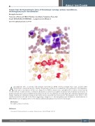
Images from the Haematologica Atlas of Hematologic Cytology: primary myelofibrosis, micromegakaryocytic transformation
Rosangela Invernizzi1,2
1University of Pavia and 2IRCCS Policlinico San Matteo Foundation, Pavia, Italy E-mail: ROSANGELA INVERNIZZI - rosangela.invernizzi@unipv.it
doi:10.3324/haematol.2021.279315
Approximately 15% of patients with primary myelofibrosis (PMF) develop terminal blast crisis, usually either myeloblastic or myelomonocytic, whereas the presence in the blood of immature cells exclusively of the megakary- ocytic type is uncommon. In this case, diagnosed as PMF for many years, a peripheral blood smear reveals a group of cells with an eccentric nucleus, condensed chromatin, no nucleoli, basophilic cytoplasm and cytoplasmic protrusions. A granulocyte precursor and an undifferentiated blast can also be seen. Platelets are often giant and sometimes agranular (top image). Immunocytochemistry using an anti-CD61 monoclonal antibody demonstrates the megakaryocytic nature of the small mononuclear cells and of the blasts suggesting a diagnosis of micromegakaryocytic leukemia as transformation of PMF. Platelets are strongly positive to the immunoalkaline-phosphatase reaction (bottom image).1
Disclosures
No conflicts of interest to disclose.
Reference
1. Invernizzi R. Myeloproliferative neoplasms. Haematologica. 2020;105(Suppl 1):49-59.
ABOUT THE COVER
haematologica | 2021; 106(9)
2297
