
Page 38 - 2021_09-Haematologica-web
P. 38
E. Onecha et al.
cases by several leukemic clones participating in the leukemic process,7 and even by genetically distinct clones segregating or combining, providing more tumor diversity.8,9 Consequently, the predominating clone at diagnosis may differ from the clone predominating in states of relapse or refractoriness.5
Previous reports on the origin and evolution of genomic mutations in AML suggest that the majority are random events that arose in hematopoietic stem/progenitor cells before they acquired the driver mutation. Patients with clon- al hematopoiesis frequently present with mutations in the genes TET2, RUNX1 and EZH2, whereas patients without clonal hematopoiesis are associated with mutations in NPM1 and FLT3.10 Furthermore, one-third of all patients with myelodysplastic syndromes (MDS) evolve to AML through a process of clonal evolution involving mutations in several genes including NPM1, RUNX1, TP53 and NRAS.11
The reappearance of leukemic disease after relapse can be through several distinct mechanisms: i) the founding clone acquires new mutations, expands and emerges as the pre- dominant clone at relapse; ii) a non-founding clone or sub- clone resists chemotherapy, acquires new mutations, expands and becomes the predominant clone in relapse; iii) an ancestral, pre-diagnostic clone evolves and emerges as the major clone at relapse; and iv) the treatment triggers the appearance of a new clone, not previously present, and gen- erates a second pathology (not a relapse per se).12,13
In a very recent study examining a cohort of adult patients with AML with NPM1 mutated at diagnosis, common can- cer pathways such as MAPK and WNT were found to be enriched in relapsed samples with loss of the NPM1 muta- tion, whereas MYC and SCF-KIT signaling pathways were enriched in relapsed samples with persistent NPM1 muta- tion.14 Similarly, in an examination of the genetic mecha- nisms of primary chemotherapy resistance in pediatric AML, mutations in FRMD8, DHX32, PIK3R1, SHANK3, MKLN1, WT1 and TP53 were maintained or even enriched in refractory disease with respect to diagnosis, and muta- tions in FLT3, PTPN11 and NRAS genes were eradicated.15
In the present study, we investigated the clinical impact of the molecular evolution of AML in patients after standard induction treatment. We performed genetic mutational stud- ies along the follow-up in refractory and relapsed AML using targeted next-generation sequencing (NGS) with a 32-gene panel. This approach involved a complete analysis of paired samples at the time of diagnosis versus refractoriness and ver- sus relapse from 23 patients who failed induction chemotherapy and/or who relapsed after CR.
Methods
Patients
The NGS-based mutational dynamics study was performed in a cohort of 23 AML patients with therapeutic failure, refractory to induction treatment (n=8), relapsed after reaching CR (n=13), and first refractory then relapsed (n=2), diagnosed between 2007 and 2015 in the Hospital 12 de Octubre, Madrid. Patients were selected from a previous sequencing study at diagnosis (n=190).16,17 The study evaluated 91 samples in total from the 23 cases at different time points: diagnosis (n=23), CR (n=31), partial remission (n=3), primary refractoriness (n=13), second-line refractoriness (n=4) and relapse (n=17). The median age at diagnosis was 59 ( range, 24–77) years and patients were treated with cytarabine and idarubicin (3+7 scheme, n=21) or with or in FLUGAZA clinical trial (azacytidine
arm, n=2) as induction treatment according to PETHEMA (Programa Español de Tratamientos en Hematología) protocols. Other clinical char- acteristics are summarized in Table 1. The study was conducted according to the Spanish law 14/2007 on biomedical research and was approved by the research ethics board of each participating institution. All patients provided informed consent.
Mutational profile workflow
DNA was extracted using Maxwell® 16 MDx (Promega Biotech Iberica SL, Madrid, Spain) and quantified on a Qubit® 2.0 Fluorometer (Invitrogen, Thermo Fisher Scientific Inc., Waltham, MA). The sequencing workflow was done with a custom NGS myeloid panel of 32 genes frequently mutated in myeloid diseases (Online Supplementary Table S1). In addition, the detection and quan- tification of mutated NPM1 sequences was performed by allele specific quantitative polymerase chain reaction (qPCR), as previ- ously described,17 using RNA as biological sample and ABL1 as the expression reference gene for normalization.18 Internal tandem duplications in FLT3 were detected using GENSCAN.19 Fastq files were processed and genomic variants were detected using RUbioSeq3.8.20 which filtering and prioritization are detailed in OnlineSupplementaryFigureS2.
Statistical analysis
All analyses were performed using the R environment (v3.4.4) for statistical computing. Fisher’s exact test was used to determine dif- ferences between two categorical variables. The median follow-up time was 18.5 (range, 2.8–127) months.
Primary refractory AML was defined as the failure to achieve CR after the first cycle of induction treatment. Partial response AML was defined as having 5–19% blast cells in bone marrow with
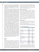
Table 1. Clinical description of patients. Patients (n=23)
Sex
Age at diagnosis Blasts at diagnosis WBC at diagnosis AML type
Karyotype at diagnosis
Cytogenetics Risk Group (ELN-2010)
HSCT
Induction therapy
Male
13 (57%)
Years, median
(range)
%, median
(range)
109/L, median
(range)
De novo
t-AML
Normal
Altered
Low
Intermediate
High
Autologous
Allogenic
Not done
3+7 scheme*
Azacytidine**
Female
10 (43%)
59
(24–78)
70
(8–06)
8.7
(1.2–145)
20 (87%)
3 (13%)
7 (30%)
16 (70 %)
3 (13%)
12 (52%)
8 (35%)
2 (9 %)
9 (39%)
12 (52%)
21 (91%)
2 (9%)
The table represents clinical data of patients included in the NGS study. WBC: white blood cells; t-AML: secondary AML to other chemotherapy; HSCT: hematopoietic stem cell transplantation. *3+7 regimen of chemotherapy: one or two induction cycles of cytarabine and idarubicin during seven and three days, respectively; and two or three consolidation cycles at high doses of cytarabine, twice a day for three alternate days followedbyallogenic-orautologous-HSCT.**Azacytidinescheme(azacytidinedays 1 to 7).
2326
haematologica | 2021; 106(9)
