
Page 230 - 2021_09-Haematologica-web
P. 230
Letters to the Editor
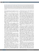
Figure 2. BCL-XL overexpression sequesters BIM and BAK and impairs S63845 mediated apoptosis induction. (A) Immunoblot analysis of the indicated BCL-2 family members was performed 24 hours (h) post-treatment initiation. One representative experiment of three independent biological replicates is shown. (B) BAK activation in MM.1S was determined by staining with antibodies against its active form. (C) MM.1S cells transduced with pcW57.1 EGFP (left panel) or pcW57.1 BCL-XL (right panel) were treated for 24 h with 0.5 μg/mL doxycycline to induce protein overexpression and afterwards exposed to the indicated treat- ments. Apoptotic cells were assessed 24 h post- treatment induction. Results indicate the mean +/- standard deviation of the mean (SDM) of three independent experiments. Differences between groups were calculated with one-way ANOVA, corrected for multiple comparison with Bonferroni-Holm correction with ****P<0.0001, **P<0.001 and *P<0.05. (D and G) Co-immunoprecipitation experiments in MM.1S cells transduced with either pcW57.1-EGFP (right panels) or pcW57.1-BCL-XL (left panels) were performed after 24 h pretreatment with 0.5 μg/mL doxycycline to induce protein overexpression and subsequent drug expo- sure for 24 h.
deeper understanding of the mechanism of activity of individual drugs to enable the optimal selection of com- bination partners and treatment sequences in clinical practice.1
In MM, the anti-apoptotic BH3 family member MCL- 1 was shown to act as a master regulator of cell survival and resistance to therapy.2,3 Accordingly, several MCL-1 inhibitors, such as S63845, S64315, AMG176 and AZD5991 are currently under evaluation in clinical tri- als.4 However, elevated expression of either BCL-XL or BCL-2 may affect the activity of MCL-1 inhibitors (MCL-1i).5 This suggests that the simultaneous targeting of multiple anti-apoptotic proteins might significantly enhance the activity of BH3 mimetics and overcome intrinsic as well as acquired drug resistance. In this con- text, a deregulation of BH3 protein family members by HDAC inhibitors (HDACi) was reported in MM6 mak- ing these compounds attractive combination partners for BH3 mimetics.
Here, we aimed to evaluate synergistic or additive combination approaches for selected BH3 mimetics. We assessed the activity of the pan-HDACi panobinostat or HDAC6i ricolinostat in combination with inhibitors tar- geting either BCL-2 (venetoclax), BCL-XL (A-1331952) or MCL-1 (S63845) in a panel of MM cell lines (MM.1S, KMS-12-BM, MOLP-8, U266, SKMM-1, RPMI-8226, OPM-2, NCI-H929). Interestingly, in six of eight MM cell lines (KMS-12-BM, MM.1S, U266, MOLP-8, NCI- H929, OPM-2) a synergistic or additive effect was observed when combining S63845+HDACi (Figure 1A to D; Online Supplementary Figure S1A to H). The combi- nation of either venetoclax or A-1331952 with HDACi led to synergistic or additive activity in three and four cell lines, respectively (Figure 1B and E; Online Supplementary Figure S1I to N).
The observed synergism was confirmed with alterna- tive MCL-1 inhibitors (AZD5991, AMG-701) (data not shown) and translated into a significant increase in apop- tosis in MM.1S, U266 and KMS-12-BM monoculture experiments using non-lethal concentrations of S63845, panobinostat and ricolinostat (Figure 1G to I). Similar effects were observed in co-culture experiments with MSCT+ stromal cells (Figure 1J to L).
Additional validation experiments confirmed the observed augmentation of the apoptotic signaling cas- cade including an enhanced release of cytochrome c (Figure 1M), cleavage of caspase 3 and PARP (Figure 1N; Online Supplementary Figure S1O and P) in all cell lines analyzed. No cell cycle alterations were observed upon single agent or combinational therapy (Online Supplementary Figure S2C to E).
The combination of S63845+HDACi proved to be particularly pronounced in the BCL-XL (co)-dependent MM cells MM1.S and U266,3,7 which otherwise did either not respond at all or only barley responded to sin- gle-agent MCL-1 inhibition.8 Moreover, BCL-XL is not only a major driver of intrinsic but also acquired MCL-1 inhibitor resistance,9 as well as dual MCL1/BCL2 inhibi- tion.10 Hence, concurrent BCL-XL inhibition seems to
optimally augment the efficacy of MCL-1 inhibitors, but prior clinical trials aiming to directly inhibit BCL-XL failed due to untoward toxicity.11,12
Based on these results we evaluated whether deregu- lation of pro- or anti-apoptotic BCL-2 family proteins by HDAC inhibitors explains the observed synergism. Single-agent treatment with S63845 monotherapy led to the accumulation of MCL-1 and BCL-XL in MM.1S (Figure 2A) and U266 (Online Supplementary Figure S2A), but not in KMS-12-BM cells (Online Supplementary Figure S2B). Conversely, combined MCL-1+HDAC inhibition led to the downregulation of BCL-XL and MCL-1 pro- tein levels in all tested cell lines prior to the onset of apoptosis (Figure 2B; Online Supplementary Figure S2A and B). In addition, a significant increase in BAK activa- tion in KMS-12-BM cells (Online Supplementary Figure S1R) was noted. In MM.1S and U266 cell lines BAK is already fully activated by S63845 alone and the combi- nation of MCL-1i and HDACi did not augment it any further (Figure 2B; Online Supplementary Figure S1M to Q), suggesting that active BAK is kept under control by alternative anti-apoptotic family members – most likely BCL-XL.
In order to test our assumption that BCL-XL inhibits apoptosis induction by S63845, we transduced MM cell lines with the Tet-on pcW57.1 vector harboring either full length BCL-XL or EGFP (control) and assessed apop- totic cells via flow cytometry. In MM.1S-BCL-XL cells, apoptosis significantly decreased upon treatment with S63845+HDACi (left panel of Figure 2C) compared to MM.1S-EGFP cells (right panel of Figure 2C). Similar findings were obtained in U266 and KMS-12-BM cells (Online Supplementary Figure 2F and G). In order to explore this rescue mechanism further, we examined the binding kinetics of BAK and BIM to BCL-XL via co- immunoprecipitations. Upon doxycycline-mediated induction of BCL-XL or EGFP protein, we treated the cells for 24 hours (h) with S63845 alone or in combina- tion with either panobinostat or ricolinostat. Strikingly, BCL-XL overexpression in S63845+HDACi-treated cells resulted in an increased association of BCL-XL and BAK as well as BIM in all investigated cell lines (left panel of Figure 2D to G; Online Supplementary Figures 2H and I and S3A to D). On the contrary, in EGFP expressing cells, BIM and/or BAK binding to BCL-XL strongly decreased upon combined S63845+HDACi exposure as compared to S63845 treatment alone (right panel of Figure 2D to G; Online Supplementary Figures S2H and I and S3A to D).
Noteworthy, we also observed cell line-specific and combination-specific effects such as an exclusive impact of ricolinostat or panobinostat on BIM-BCL-XL, but not BAK-BCL-XL binding, in MM.1S-EGFP and U266-EGFP cells, respectively (right panel of Figure 2F and G; right panel of Online Supplementary Figure 2H and I). Furthermore, in KMS-12-BM-EGFP cells BIM was not associated with BCL-XL (right panel of Online Supplementary Figure S3C), as BIM is rather sequestered by BCL-2 (data not shown) in line with the MCL-1/BCL-
2518
haematologica | 2021; 106(9)
