
Page 185 - 2021_09-Haematologica-web
P. 185
Interleukin-1 receptor in sickle cell disease
Results from this study support a critical role of non- hematopoietic IL-1R signaling in mediating acute brain tis- sue damage in SCD mice in to the setting of ischemic stroke. This effect was associated with IL-1R-mediated regulation of endothelial adhesive properties. Signaling via the endothelial IL-1R leads to upregulation of endothelial adhesion molecules with resultant increases in leukocyte- endothelial interactions and tissue leukocyte infiltration.40 This signaling pathway involves enhanced NFκB signal- ing.40 Enhanced endothelial expression of adhesion mole- cules is detrimental in SCD, promoting vascular occlu-
sions and pain crises.52 Regulation of endothelial IL-1R responses to IL-1β has also been shown to occur indirectly by leukocyte interactions with selectins. For example, mice with leukocyte deficiency of p-selectin glycoprotein ligand-1 (Psgl-1) are resistant to IL-1β-mediated stimula- tion of endothelial adhesion molecule expression and show reduced leukocyte-endothelial interactions.40 This is potentially relevant to SCD as an antibody to p-selectin, crizanlizumab, has been shown in human clinical trials to reduce the frequency of vaso-occlusive events.53 Preclincal studies have also shown that SCD mice treated with an
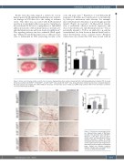
AB
CD
Figure 2. Stroke area following middle cerebral artery occlusion. Representative brain sections stained with 4% 2,3,5-triphenyltetrazolium chloride (TTC) to assess stroke size (in white) of (A) Wt,WTbmt, (B) Wt,SCDbmt, (C) IL1R-/-,SCDbmt mice, and (D) IL6-/-,SCDbmt mice. (E) Quantification of stroke volume (mean ± standard deviation). The brain sections were imaged with a Nikon SMZ-2T microscope and Spot Idea camera model 29.2-13MP using at Nikon 0.45x TV lens and Spot 5.1 software. *P<0.05 as determined by ANOVA.
E
Figure 3. Post-stroke macrophage infiltra- tion. Representative images of MAC3-posi- tive cells in the peri-infact area of Wt,WTbmt, Wt,SCDbmt, IL1R-/-,SCDbmt and IL6-/-,SCDbmt mouse brains, and quantification (mean ± standard deviation). A Nikon SE upright microscope and a Nikon DS-Fi3 camera was used to capture 10x and 20x images of ipsi- lateral brain and 10x images of contralateral brain. Dotted line denotes transition between infarcted area and heathy tissue. *P<0.05 as determined by ANOVA.
haematologica | 2021; 106(9)
2473
