
Page 9 - 2021_06-Haematologica-web
P. 9
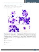
Images from the Haematologica Atlas of Hematologic Cytology: visceral leishmaniasis
Rosangela Invernizzi1 and Antonello Malfitano2
1University of Pavia and 2IRCCS Policlinico San Matteo Foundation, Pavia, Italy E-mail: ROSANGELA INVERNIZZI - rosangela.invernizzi@unipv.it
doi:10.3324/haematol.2021.279161
Bone marrow smears remain the diagnostic gold standard for visceral leishmaniasis, although Donovan’s bodies, the identifying marker of the disease, may also be found in lymph nodes as well as in spleen fine-needle aspirate smears. These bodies are the asexual form of the parasite, the so-called amastigotes. They are oval, measure 1-5 μm long by 1-2 mm wide, and show a round nucleus and a bar-shaped inclusion, the kinetoplast, which is the remnant of the flagellum of the promastigote. Amastigotes of Leishmania donovani may be numerous inside bone marrow macrophages (Figure A-D) or extracellularly as macrophages can be broken during preparation of the smear (Figure E). They may also be observed in the peripheral blood within the cytoplasm of granulocytes, particularly after centrifugation and concentration of white blood cells (Figure F).1
Disclosures
No conflicts of interest to disclose.
Contributions
Both authors contributed.
Reference
1. Malfitano A, Invernizzi R. Parasitic and fungal diseases. Haematologica. 2020;105(Suppl 1):29-39.
ABOUT THE COVER
haematologica | 2021; 106(8)
2037
