
Page 235 - 2021_06-Haematologica-web
P. 235
Letters to the Editor
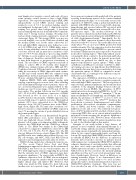
nant lymphocytes (negative control) and renal cell carci- noma (positive control; known to have a high CRM1 expression).6 Two expert hematopathologists (RLK, AJW) independently scored CRM1 nuclear staining and assigned a score of 0-3; 0 (no nuclear staining; equal to non-malignant background cells), 1 (dim/weak nuclear staining but greater than background), 2 (moderate nuclear staining with nuclear detail still visible behind the stain) and 3 (strong nuclear staining obscuring most nuclear detail; staining intensity equivalent to renal cell carcinoma), Figure 1A. The average CRM1 score per case across all available cores on the TMA was calculated and the median CRM1 score for the entire cohort was 2.5. Low and high-CRM1 expression were defined as scores of 0-2.4 (CRM1-low) and 2.5-3.0 (CRM1-high), respec- tively. Scoring reliability between reviewers and between cores was assessed and an intra-class correlation coeffi- cient of 0.75-0.90 was defined as “good scoring reliabili- ty”. All time-to-event analyses were conducted from the time of diagnosis. Event-free survival (EFS) was defined as time from diagnosis to progression, retreatment, or death. The association of CRM1 expression and risk of failing to achieve EFS at 24 months after diagnosis (EFS24) was estimated using odds ratios (OR) and 95% Confidence Intervals (CI) from logistic regression models, while the association of CRM1 expression with continu- ous EFS and overall survival (OS) was estimated using Kaplan-Meier method and hazard ratios (HR) and 95% CI were calculated through Cox regression models.
Eighteen DLBCL TMA with optimal overlap with ongoing sequencing projects were used. After excluding non-chemoimmunotherapy treated patients, tumor tis- sue cores from 282 patients were analyzed (Table 1). The median age was 61 years (range, 18-93) and 166 (59%) were males. The median follow-up was 88.6 months (95% CI: 82.9-95.5). Quantitative expression of CRM1 was detected in 99% of cases (score of 1 or higher, n=278); therefore the intensity of CRM1 expression was assessed with respect to patient outcomes. One-hundred patients (35%) were categorized in to the CRM1-low intensity cohort; 182 (65%) patients were in the CRM1- high cohort, (Figure 1B). The intra-class correlation coef- ficient to measure scoring reliability of CRM1 expression was 0.79, meeting the criteria of good reliability between the hematopathologists.
There were no differences in International Prognostic Index (IPI), performance score, lactate dehydrogenase level, age, high-risk disease (double or triple-hit lym- phoma), cell-of-origin, or treatment modality at diagnosis between the CRM1-high and the CRM1-low cohorts, (Table 1). The EFS24 was similar in the CRM1-high cohort (30%) compared to CRM1-low cohort (27%), OR 1.09, 95% CI: 0.73-1.63; P=0.67, (Figure 1C). The OS was not different between cohorts (Figure 1D). The EFS and OS results were similar when adjusted for age, sex, IPI, cell-of-origin, and MYC, BCL2 and BCL6 protein expression and rearrangement status (data not shown).
CRM1 is being exploited as a therapeutic target in can- cer.1 The recent United States Food and Drug Administration approval of selinexor for the treatment of R/R DLBCL was based on an overall response rate of 28% with single-agent selinexor suggesting the potential that the intensity level of CRM1 expression may have prognostic significance.5 In a prior study in DLBCL, the qualitative expression level of CRM1 was an independ- ent negative prognostic marker which associated with higher clinical stage and inferior OS.7 However, that study was small (n=131), limited by short follow-up (median was not reported; range, 14-65 months), and
heterogeneous treatments with nearly half of the patients receiving chemotherapy instead of the current standard of chemoimmunotherapy.8 A second study assessed the expression of CRM1 by using a polyclonal-antibody in patients with DLBCL who were treated with chemoim- munotherapy and reported inferior OS associated with high-level of CRM1 expression in activated B-cell or dou- ble-expresser types.9 The median follow-up of the patients was not mentioned in that study and the OS was lower than typical real-world patients with DLBCL treat- ed with chemoimmunotherapy.10 Importantly, the fre- quency of CRM1 expression in tumor cells in that study was low at only 40% of cases. This result differs from our study where 99% of cases were CRM1 positive but with variable intensity. The low expression level in that study is surprising given that CRM1/XPO1 is a protein critical for normal cellular function and is expected to be present in all cells to varying degrees.11 Our study utilized a mon- oclonal-antibody in contrast to the polyclonal-antibodies used in the two prior studies. In general, monoclonal- antibodies are preferred for clinical use due to their improved specificity for a given epitope.12 While techni- cal differences in IHC protocols may contribute to differ- ent staining outcomes, we hypothesize that CRM1 poly- clonal-antibodies may have lower sensitivity in detecting the particular epitope of CRM1 as compared to mono- clonal-antibodies, accounting for the differences in posi- tivity rates across DLBCL.
The expression of CRM1 in solid tumors has also been assessed in the past and shown a negative prognostic value with high CRM1 expression.2 The heterogeneity of tumor biology, patient populations, and treatments may contribute to the disparate findings between solid can- cers and ours here in DLBCL. The prevalence of XPO1 mutation has been documented in DLBCL to be 2-4%, in which the majority were XPO1E571K mutation.13,14 In pri- mary mediastinal B-cell lymphoma and Hodgkin lym- phoma, the prevalence of XPO1E571K is close to 25% in some studies.15 Moreover, the XPO1E571K mutation has been shown to promote lymphomagenesis and cellular proliferation, alter nuclear cytoplasmic compartmenta- tion of CRM1, and better sensitize cells for CRM1 inhibitors in vitro and in vivo.1 The relatively low preva- lence of XPO1 mutation in LCL, and lack of whole exome sequencing (WES) data on our current patient population prevented us from assessing the prognostic significance of XPO1 mutations in LGL. However, future studies that include WES data on large LCL patient populations may be able to shed further light on this matter.
Strengths of our study include a large dataset, all patients received standard chemoimmunotherapy in a real-world setting, uniform assessment of CRM1 expres- sion using a monoclonal-antibody, independent interpre- tation by two expert hematopathologists, and long fol- low-up. We report that CRM1/XPO1 protein is expressed in virtually all DLBCL but the difference in expression intensity did not predict outcomes in patients treated with regimens not containing the CRM1/XPO1-inhibitor, selinexor. Limitations of our study include that, although we have used easily reproducible methods to assess CRM1 expression in tumor cells with good scoring relia- bility between the two reviewers, the intensity grading algorithm used has not been tested among larger num- bers of pathologists. However, based on differences in staining intensities (Figure 1) we demonstrated, there may be potential for this assay to be used in future prospective trials to learn if intensity predicts response to CRM1/XPO1-inhibitor treatment. This issue has not been described in any of the clinical trials which have
haematologica | 2021; 106(8)
2263
