
Page 220 - 2021_06-Haematologica-web
P. 220
Letters to the Editor
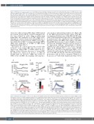
Figure 1. TRPV2 protein in mouse red blood cells. (A, B) Western blot of wild-type, Trpv2 knockout (KO) (A) and Trpc6 KO (B) red blood cell (RBC) proteins, using antibodies against mouse TRPV2, TRPC6 and b-actin. (C) Semi-quantitative analysis of differentially expressed proteins identified by mass spectrometry in wild- type and Trpv2 KO RBC lysates. Up- and down-regulated proteins were identified based on at least 2-fold changes with a P-value <0.05, calculated by an unpaired two-tailed Student t test. The heatmap shows the Z-scores of the exponentially modified protein abundance index (emPAI) values of mass spectrometry measurements of five independent wild-type and four independent Trpv2 KO samples. (D) Relative abundance of the TRPV2 protein compared to that of the 1,450 proteins identified in mouse erythrocyte membrane fractions. Rank represents the order of the identified protein obtained by spectral counting with the P-value calculated by an unpaired Student t test. NA, not applicable. (E) Hematologic parameters of the blood from wild-type and Trpv2 KO. RBC: red blood cell; HGB: hemoglobin; HCT: hematocrit; MCV: mean corpuscular volume; MCH: mean corpuscular hemoglobin; MCHC: mean corpuscular hemoglobin concentration; RDW: red cell distribution width with P-value calculated by an unpaired two-tailed Student t test. (F) Hemolysis (%) of RBC collected from wild-type (black) and Trpv2 KO mice (red) in buffer A (149 mM NaCl, 2 mM CaCl2, 4 mM KCl, 2 mM HEPES, pH7.4), diluted 26-fold in buffer B (0-149 mM NaCl, 2 mM HEPES, pH 7.4) as indicated; extracellular [Ca2+] was kept at ~76 mM. (G) Tonicity at which 50% lysis occurred (C50), calculated by sigmoidal fitting from experiments in (F). Single values and mean ± standard error of mean from five independent experiments performed in triplicate are shown with the P-value calculated by an unpaired two- tailed Student t test.
elicited Ca2+ influx in human RBC (Figure 3B-D). Inward and outward currents with the outward rectifying IV rela- tion were obtained by patch clamp recordings from human RBC after application of Δ9-THC (Figure 3E-G). Although the currents obtained from human RBC have a small amplitude, their IV match the TRPV2 current signa- ture obtained from COS-7 overexpressing human TRPV2 cDNA upon application of Δ9-THC or CBD (Online Supplementary Figure S3B, C).
Assessment by confocal microscopy revealed that 95.3±2.4% of the human RBC had a biconcave disc- shaped form. Adding CBD or Δ9-THC shifted the mor- phology of these biconcave discocytes to concave RBC, the stomatocytes, which in the presence of CBD and Δ9-THC make up 92.7±1.3% (CBD) and 66.3±17.1% (THC) of the total RBC, the remaining cells being disco-
cytes and more spherical-shaped spherocytes (Figure 3H,
I). The TRPV2 agonist-induced shape change of the RBC
was maintained in the presence of the CB1 and CB2 antag-
onists (Online Supplementary Figure S3D, E), indicating that
the major fraction of the cannabinoids’ effect on RBC mor-
phology is mediated by TRPV2. After addition of Δ9-THC,
human RBC showed reduced osmotic fragility, as demon-
strated by the left-shifted hemolysis curve in response to
the hypotonicity challenge, independently of whether
cannabinoid receptor antagonists were absent or present
(Figure 3J, K). Similarly, but to a lesser extent, Δ9-THC
shifts the C50 value after treating WT murine RBC to lower
tonicities (C , in the absence, 49.05±1.53, and in the pres- 50
ence of Δ9-THC, 46.08±1.55). This effect was reversed by pretreatment with the KCa3.1 antagonist TRAM-34, in the presence of 76 mM (Online Supplementary Figure S3F, G)
ABCD
EFGH
2248
Figure 2. TRPV2 function in mouse red blood cells. (A-D) In- and outward currents at -80 and +80 mV shown as mean ± standard error of mean (SEM), recorded from mouse red blood cells (RBC) using a patch pipette (A) or a miniaturized patch clamp system (port-a-patch) (C) plotted versus time (number of cells in brack- ets). TRPV2 currents were activated by the application of 2-APB (black line) in the absence and presence of 10 mM ruthenium red (RR, blue line) with the cor- responding current-voltage relationships (IV) at the peak net currents (Imax net), shown as mean ± SEM in (B) and (D). Patch pipette resistances were 10 - 15 MΩ when filled with standard internal solution (in mM): 120 Cs-glutamate, 8 NaCl, 1 MgCl2, 10 HEPES, 10 1,2-bis(2-aminophenoxy)ethane-N,N,N',N'-tetraacetic acid tetracesium salt (Cs-BAPTA), 3.1 CaCl2 (100 nM free Ca2+, calculated with WebMaxC), pH7.2 with CsOH. Standard external solution contained (in mM): 140 NaCl, 2 MgCl2, 1 CaCl2, 10 HEPES, 10 glucose, pH 7.2 with NaOH. For experiments with the miniaturized patch system, the intracellular solution contained (in mM): 60 Cs-methansulfonate, 8 NaCl, 1 MgCl2, 3.1 CaCl2, 60 CsF, 10 HEPES, 10 BAPTA (100 nM free Ca2+, calculated with WebMaxC), 10 glucose, pH 7.2 with CsOH and the extracellular solution contained (in mM): 140 NaCl, 2 MgCl2, 1.35 CaCl2, 10 HEPES, 10 glucose, pH 7.2 with NaOH. (E, G) Mean Fluo-4 fluores- cence (F/F0) traces showing changes in the cytosolic [Ca2+] of RBC isolated from wild-type (black) and Trpv2 KO mice (red) in the absence (E) and presence (G, blue) of the CB1/CB2-receptor antagonists AM251 and JTE907 (100 nM each), challenged by the application of 30 mM Δ9-tetrahydrocannabinol (Δ9-THC, line). Ca2+-imaging measurements were performed in the presence of a Tyrode solution (in mM): 135 NaCl, 5.4 KCl, 1 MgCl2, 10 HEPES, 10 glucose, and 1.8 CaCl2, pH 7.35; RBC were loaded with 5 μM Fluo-4 and the fluorescence was excited at 488 nm every 3 seconds with the emitted fluorescence detected at >515 nm. (F, H) Summary of peak amplitudes from (E) and (G) shown as mean ± SEM with P-values calculated by the unpaired two-tailed Student t test (ns, not significant). Numbers of measured cells (x) within (y) independent experiments are indicated in brackets and bars.
haematologica | 2021; 106(8)
